|
|
| (22 intermediate revisions by 6 users not shown) |
| Line 1: |
Line 1: |
| − | ==Overview==
| + | <big><center>[[Oral Cavity - Salivary Glands - Anatomy & Physiology|'''BACK TO SALIVARY GLANDS- ANATOMY & PHYSIOLOGY''']]</center></big> |
| | + | <big><center>[[Parotid|'''BACK TO PAROTID SALIVARY GLAND- ANATOMY & PHYSIOLOGY''']]</center></big> |
| | | | |
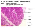
| − | [[Image:Serous Salivary Gland.jpg|thumb|right|250px|Serous Salivary Gland Histology - Copyright RVC 2008]]
| + | '''Serous Salivary Glands''' |
| − | The serous salivary gland has a connective tissue capsule and septa dividing the parenchyma into lobes. There is a duct system. Interlobular ducts run in the tissue septum lined by cuboidal to columnar epithelium.
| |
| | | | |
| − | Intralobular ducts run within the lobules. Striated intralobular ducts are lined with cuboidal epithelium. Intercalated intralobular ducts are lined with low cuboidal to simple squamous epithelium. Serous acini secrete a watery solution rich in proteins with spherical nuclei. Cells are pyramidal, cuboidal or crescent shaped.
| + | [[Image:Serous Salivary Gland.jpg|thumb|right|150px|Serous Salivary Gland Histology - Copywright RVC 2008]] |
| − | <br>
| |
| − | {{Template:Learning
| |
| − | |flashcards= [[Salivary Gland Anatomy & Physiology Flashcards|Salivary Glands Anatomy & Physiology Flashcards]]
| |
| − | |powerpoints= [[Oral Cavity Histology resource|Oral Cavity Histology, see part 2 for salivary glands]] | |
| − | |Vetstream = [https://www.vetstream.com/canis/search?s=salivary Salivary Gland Diseases] | |
| − | | |
| − | }}
| |
| − | | |
| − | ==Webinars==
| |
| − | <rss max="10" highlight="none">https://www.thewebinarvet.com/dentistry/webinars/feed</rss>
| |
| − | | |
| − | [[Category:Salivary Glands - Anatomy & Physiology]]
| |
| − | [[Category:A&P Done]]
| |
