Difference between revisions of "Proventriculus - Anatomy & Physiology"
Jump to navigation
Jump to search
| Line 33: | Line 33: | ||
==Histology== | ==Histology== | ||
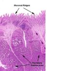
| + | [[Image:Proventriculus Histology.jpg|thumb|right|150px|Proventriculus Histology - Dr. Thomas Caceci and Dr. Ihab El-Zhogby, Department of Histology, Faculty of Veterinary Medicine, Zagazig University, Egypt]] | ||
*Mucous cells | *Mucous cells | ||
Revision as of 15:53, 9 July 2008
Introduction
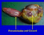
The proventriculus is also referred to as the muscular stomach. It is connected by the isthmus to the gizzard.
Structure and Function
- A storage organ in fish and flesh eating birds
- Appropriate to a soft diet
- Secretes digestive enzymes
- Contacts the left lobe of the liver ventrally and laterally
- Related dorso-caudally to the spleen
- More cranial than the gizzard
- Lies to the left of the midline of the bird
- Spindle/fusiform shaped
- Roughly 4cm long
- Lumen diameter similar to the oesophagus
Histology
- Mucous cells
- Columnar epithelium
- Basolphilic
- Papillae- through which collecting ducts from glands run
- Lamina propria run into the papillae
- Hydrochloric acid and pepsin produced
- Glands in the submucosa
- Single tubular glands are grouped into lobules with a common opening into a papillae
- Serous membrane of mesothelial cells attached to the outer longitudinal layer of muscle
- 3 layers of lamina muscularis
- No parietal cells

