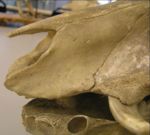
Difference between revisions of "Skull and Facial Muscles - Anatomy & Physiology"
(→Feline) |
|||
| Line 10: | Line 10: | ||
The viscerocranium is the pharyngeal skeleton. It is derived only from the neural crest and undergoes endochondral and intramembranous ossification. | The viscerocranium is the pharyngeal skeleton. It is derived only from the neural crest and undergoes endochondral and intramembranous ossification. | ||
| + | |||
| + | The shape and size of the skull varies widely, not only between species but also with age, breed and sex of similar species. | ||
==Structure== | ==Structure== | ||
| Line 35: | Line 37: | ||
===Occipital Bone=== | ===Occipital Bone=== | ||
| − | * | + | *Forms the nuchal wall |
| + | |||
| + | *Forms the foramen magnum | ||
| + | |||
| + | *Basilar part (pars basilaris) | ||
| + | **Caudal base of the cranium | ||
| + | **Rostral to foramen magnum | ||
| + | **Joined by cartilagenous suture to basisphenoid bone | ||
| + | **Muscular tubercules on ventral surface where the flexors of the head and neck attach | ||
| + | **Caudal cranial fossa enclose the pons and medulla oblongata | ||
| + | |||
| + | *Squamous part (pars squamosa) | ||
| + | **Dorsal to lateral parts and occipital condyles | ||
| + | **Nuchal crest present | ||
| + | ***Easily palpable | ||
| + | ***Landmark for collection of cerebrospinal fluid (CSF) | ||
| + | **External occipital protuberances present which provide muscle attachment sites for the nuchal ligament | ||
| + | |||
| + | *Lateral parts (partes laterales) | ||
| + | **Lateral borders of foramen magnum | ||
| + | **Occipital condyles present which articulate with the atlas to form the atlanto-occipital joint | ||
| + | **Paracondylar process present which provides muscle attachment sights for muscles of the head | ||
| + | **Hypoglossal canal | ||
===Sphenoid Bone=== | ===Sphenoid Bone=== | ||
| Line 66: | Line 90: | ||
===Mandible=== | ===Mandible=== | ||
| + | |||
| + | ==Foramen== | ||
| + | |||
| + | *Jugular Foramen | ||
| + | **Located either side of basilar part of occipital bone | ||
| + | **Adjacent to tympanic bulla | ||
| + | |||
| + | *Foramen Magnum | ||
| + | **Formed by occipital bones | ||
| + | |||
| + | *Hypoglossal Canal | ||
| + | **Between paracondylar and condylar processes on lateral part of occipital bone | ||
==Species Differences== | ==Species Differences== | ||
| Line 76: | Line 112: | ||
*2 halves of the mandible do not fuse allowing some movement | *2 halves of the mandible do not fuse allowing some movement | ||
| + | |||
| + | *External sagittal crest arises from nuchal crest | ||
[[Image:Lion skull.jpg|thumb|right|150px|Feline skull (Lion) - Copyright RVC]] | [[Image:Lion skull.jpg|thumb|right|150px|Feline skull (Lion) - Copyright RVC]] | ||
| Line 87: | Line 125: | ||
*2 halves of the mandible do not fuse allowing some movement | *2 halves of the mandible do not fuse allowing some movement | ||
| − | *Weak sagittal crest | + | *Weak external sagittal crest arises from nuchal crest |
[[Image:Horse Skeleton.jpg|thumb|right|150px|Horse Skeleton - Copyright Nottingham]] | [[Image:Horse Skeleton.jpg|thumb|right|150px|Horse Skeleton - Copyright Nottingham]] | ||
===Equine=== | ===Equine=== | ||
| − | *Weak sagittal crest | + | |
| + | *Weak external sagittal crest arises from nuchal crest | ||
*Long skull length | *Long skull length | ||
| Line 111: | Line 150: | ||
*[[Anatomy & Physiology of the Horn|Cornual]] process on frontal bone | *[[Anatomy & Physiology of the Horn|Cornual]] process on frontal bone | ||
| + | |||
| + | *Nuchal crest reduced to nuchal line | ||
*Prominent temporal line | *Prominent temporal line | ||
Revision as of 12:27, 30 July 2008
Introduction
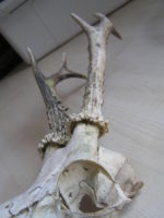
The skull is divided into three components- the neurocranium, the dermatocranium and the viscerocranium. The skull also includes the hyoid apparatus, mandible, ossicles of the middle ear and the cartilage of the larynx, nose and ear. The skull protects the brain and head against injury and supports the structures of the face. In some animals the skull is also used for defensive actions, for example in horned ungulates such as red deer stags.
The neurocranium develops from the neural crest and mesoderm and undergoes endochondral ossification. It lies ventral to the brain.
The dermatocranium lies dorsal to the brain and develops from the neural crest and mesoderm. It undergoes intramembranous ossification.
The viscerocranium is the pharyngeal skeleton. It is derived only from the neural crest and undergoes endochondral and intramembranous ossification.
The shape and size of the skull varies widely, not only between species but also with age, breed and sex of similar species.
Structure
- The skull is made of many smaller bones
- Most of the skull bones are paired
- Cartilage or fibrous tissue separates the bones of the skull in the young animal
- Once growth has ceased, the sutures begin to ossify
Function
- Protection of brain
- Support facial muscles by providing origin and insertion sites
- Foramen provide entry and exit places for the vasculature and nervous system
- Defense
Bones of the Skull
Occipital Bone
- Forms the nuchal wall
- Forms the foramen magnum
- Basilar part (pars basilaris)
- Caudal base of the cranium
- Rostral to foramen magnum
- Joined by cartilagenous suture to basisphenoid bone
- Muscular tubercules on ventral surface where the flexors of the head and neck attach
- Caudal cranial fossa enclose the pons and medulla oblongata
- Squamous part (pars squamosa)
- Dorsal to lateral parts and occipital condyles
- Nuchal crest present
- Easily palpable
- Landmark for collection of cerebrospinal fluid (CSF)
- External occipital protuberances present which provide muscle attachment sites for the nuchal ligament
- Lateral parts (partes laterales)
- Lateral borders of foramen magnum
- Occipital condyles present which articulate with the atlas to form the atlanto-occipital joint
- Paracondylar process present which provides muscle attachment sights for muscles of the head
- Hypoglossal canal
Sphenoid Bone
Temporal Bone
Frontal Bone
Parietal Bone
Ethmoid Bone
Nasal Bone
Lacrimal Bone
Zygomatic Bone
Maxilla
Incisive Bone
Palatine Bone
Vomer
Pterygoid Bone
Mandible
Foramen
- Jugular Foramen
- Located either side of basilar part of occipital bone
- Adjacent to tympanic bulla
- Foramen Magnum
- Formed by occipital bones
- Hypoglossal Canal
- Between paracondylar and condylar processes on lateral part of occipital bone
Species Differences
Canine
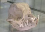
- Dogs have different skull lengths depending on breed
- Mesocephalic dogs have average conformation
- Dolichocephalic dogs have longer skull lengths
- Brachycephalic dogs have shorter skull lengths
- 2 halves of the mandible do not fuse allowing some movement
- External sagittal crest arises from nuchal crest
Feline
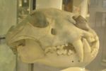
- The mandible appears globular in shape
- Large orbits with complete bony margins
- Large tympanic bulla which can be palpated
- 2 halves of the mandible do not fuse allowing some movement
- Weak external sagittal crest arises from nuchal crest
Equine
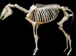
- Weak external sagittal crest arises from nuchal crest
- Long skull length
- Orbit placed more laterally with a complete bony rim
- Strong zygomatic arch which continues on to form the facial crest
- Deep nasoincisive notch
- Prominent hamular process
- Large mandible
- Vascular notch on mandible
- High ramus
Ruminant
- Skull is short and wide
- Cornual process on frontal bone
- Nuchal crest reduced to nuchal line
- Prominent temporal line
- Elevated orbital ring which is complete
- No facial crest
- Prominent tympanic bullae
- Nasoincisive notch present
Porcine
- Thick nucal crests
- Prominent temporal line
- Orbit is incomplete and small
- Strong and deep zygomatic arch
- Large tympanic bullae
- High caudal part of the skull
Avian
- Pneumatised skull bones
- Spaces in skull bones which connect to airways in the head rather than the air sacs
- Large orbits
- Skull plates are separated by spongy bone
- A single occipital condyle articulates with the atlas allowing more rotation of the head
- In parrots, the nasal bone and frontal bone are joined by a flexible cartliage structure allowing greater jaw opening which is called the craniofacial hinge. This allows kinesis to occur.
- Thin jugal arch (equivalent to zygomatic arch)
- Middle ear contains only the columella (equivalent to the stapes)
