Difference between revisions of "File:Equine protozoal myeloencephalitis.jpg"
(Sarcocystis neurona stages and lesions. (A). Cross section of spinal cord of horse with focal areas of discoloration (arrows) indicative of necrosis. Unstained. (B). Section of spinal cord of a horse with severe EPM. Necrosis, and a heavily infected neu) |
(No difference)
|
Revision as of 14:27, 21 December 2008
Sarcocystis neurona stages and lesions.
(A). Cross section of spinal cord of horse with focal areas of discoloration (arrows) indicative of necrosis. Unstained.
(B). Section of spinal cord of a horse with severe EPM. Necrosis, and a heavily infected neuron (arrows), all dots (arrows) are merozoites. H and E stain .
(C). Higher magnification of a dendrite with numerous merozoites (arrows). One extracellular merozoite (arrowhead) and a young schizont (double arrowhead).
(D). Section of brain of an experimentally-infected mouse stained with anti-S. neurona antibodies. Note numerous merozoites (arrows).
(E). Immature schizonts in cell culture. A schizont with multilobed nucleus (arrow) and a schizont with differentiating merozoites (arrowheads). Giemsa stain.
(F). Mature sarcocysts with hairlike villar protrusions (double arrowheads) on the sarcocyst wall. H and E stain.
(G). Mature live sarcocyst with numerous septa (arrows) and hairlike protrusions on the sarcocyst wall (double arrowheads). Unstained.
(H). An oocyst with two sporocysts each with banana-shaped sporozoites. Unstained.
http://commons.wikimedia.org/wiki/File:Equine_Protozoal_Myeloencephalitis.jpg
Wikimedia Commons
File history
Click on a date/time to view the file as it appeared at that time.
| Date/Time | Thumbnail | Dimensions | User | Comment | |
|---|---|---|---|---|---|
| current | 14:27, 21 December 2008 | 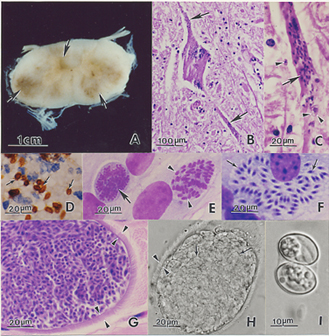 | 329 × 336 (151 KB) | Nabrown (talk | contribs) | Sarcocystis neurona stages and lesions. (A). Cross section of spinal cord of horse with focal areas of discoloration (arrows) indicative of necrosis. Unstained. (B). Section of spinal cord of a horse with severe EPM. Necrosis, and a heavily infected neu |
You cannot overwrite this file.
File usage
The following 2 files are duplicates of this file (more details):
- File:Equine Protozoal Myeloencephalitis.jpg
- File:Equine Protozoal Myeloencephalitis.jpg from Wikimedia Commons
The following 2 pages use this file: