Difference between revisions of "File:Venezuelan equine encephalitis virus.jpg"
Jump to navigation
Jump to search
(Photomicrograph of mouse brain tissue after dying of Venezuelan Encephalitis. Reveals neural necrosis and edema. ''This image is a work of the Centers for Disease Control and Prevention (CDC) and was sourced from Wikimedia Commons. Copyright of the CDC's) |
|||
| Line 1: | Line 1: | ||
| − | Photomicrograph of mouse brain tissue after dying of Venezuelan Encephalitis. Reveals neural necrosis and edema. | + | Photomicrograph of mouse brain tissue after dying of Venezuelan Encephalitis. Reveals neural necrosis and edema. This image is a work of the Centers for Disease Control and Prevention (CDC) and was sourced from Wikimedia Commons. Copyright of the CDC's Public Health Image Library (PHIL) Image #2809, 2006. |
Latest revision as of 13:09, 7 July 2010
Photomicrograph of mouse brain tissue after dying of Venezuelan Encephalitis. Reveals neural necrosis and edema. This image is a work of the Centers for Disease Control and Prevention (CDC) and was sourced from Wikimedia Commons. Copyright of the CDC's Public Health Image Library (PHIL) Image #2809, 2006.
File history
Click on a date/time to view the file as it appeared at that time.
| Date/Time | Thumbnail | Dimensions | User | Comment | |
|---|---|---|---|---|---|
| current | 13:08, 7 July 2010 | 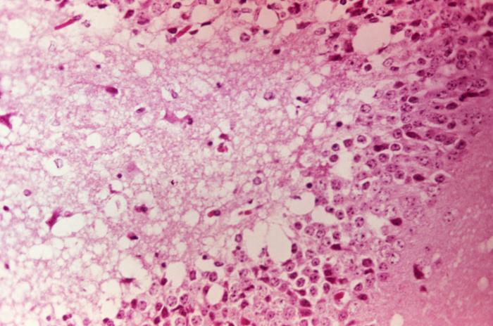 | 700 × 464 (62 KB) | Nmr28 (talk | contribs) | Photomicrograph of mouse brain tissue after dying of Venezuelan Encephalitis. Reveals neural necrosis and edema. ''This image is a work of the Centers for Disease Control and Prevention (CDC) and was sourced from Wikimedia Commons. Copyright of the CDC's |
You cannot overwrite this file.
File usage
The following file is a duplicate of this file (more details):
- File:Venezuelan equine encephalitis virus.jpg from Wikimedia Commons
The following page uses this file: