Difference between revisions of "File:Hpylori.jpg"
Jump to navigation
Jump to search
| Line 10: | Line 10: | ||
== Licensing: == | == Licensing: == | ||
| − | {{ | + | {{cc-att-2.0}} |
Latest revision as of 17:58, 25 July 2010
Summary
| Description |
Electron micrograph of H. pylori possessing multiple flagella (negative staining) |
|---|---|
| Date |
26 January 2006 |
| Source |
(Original Source: http://info.fujita-hu.ac.jp/~tsutsumi/photo/photo002-6.htm) http://commons.wikimedia.org/wiki/File:EMpylori.jpg |
| Author |
Yutaka Tsutsumi (M.D. Professor Department of Pathology Fujita Health University School of Medicine) |
| Permission (Reusing this file) |
See below |
Licensing:
| This file is licensed under the Creative Commons Attribution Non-Commercial & No Derivative Works License |
File history
Click on a date/time to view the file as it appeared at that time.
| Date/Time | Thumbnail | Dimensions | User | Comment | |
|---|---|---|---|---|---|
| current | 09:46, 8 July 2010 | 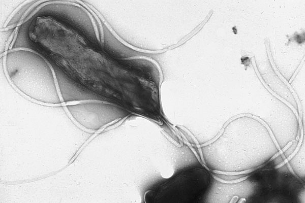 | 600 × 400 (65 KB) | Svycrnj (talk | contribs) | H.pylori - © Yutaka Tsutsumi, M.D. Professor Department of Pathology Fujita Health University School of Medicine, Wikimedia Commons. Free use copyright, taken on 8/7/2010. |
You cannot overwrite this file.
File usage
The following file is a duplicate of this file (more details):
- File:EMpylori.jpg from Wikimedia Commons
The following page uses this file: