Difference between revisions of "Category:Protozoa"
| (4 intermediate revisions by one other user not shown) | |||
| Line 5: | Line 5: | ||
Protozoa usually range from 10μm-50μm but can grow up to 1mm. Thus, they are usually observed and classified using a microscope. | Protozoa usually range from 10μm-50μm but can grow up to 1mm. Thus, they are usually observed and classified using a microscope. | ||
| − | Not all protozoa are harmful. For example, the [[ | + | Not all protozoa are harmful. For example, the [[Rumen - Anatomy & Physiology|rumen]] of ruminants and the [[Caecum - Anatomy & Physiology|caecum]] and [[Colon - Anatomy & Physiology|colon]] of horses are full of symbiotic protozoa. |
</div> | </div> | ||
| Line 40: | Line 40: | ||
| + | ==Useful Resources== | ||
| + | *http://www.veterinariavirtual.uab.es/parasito/diagnos003$/coproeq.htm | ||
| + | ''Brilliant microscopic pictures of protozoa and helminths'' | ||
| + | *http://www.vet.uga.edu/VPP/clerk/siegel/index.php | ||
| + | ''Detailed information and images, including clincial signs and pathogenesis, of East Coast Fever'' | ||
| + | |||
| + | *http://cal.vet.upenn.edu/projects/dxendopar/index.html#host | ||
| + | ''Useful online resource for diagnosing parasitic infections, courtesy of the Laboratory of Parasitology, University of Pennsylvania School of Veterinary Medicine'' | ||
| + | |||
| + | *http://www.veterinaryhub.com/giardia-in-dogs/ | ||
| + | ''Giardia infestation in dogs, common signs and symptoms, treatment and control'' | ||
Latest revision as of 19:18, 27 September 2014
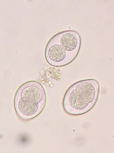
All protozoa are unicellular eukaryotic organisms which store their genetic information in chromosomes in a nuclear envelope. Protozoa are classified depending on their structure and life cycle. This reflects the similarities of the diseases which they cause. Protozoa are heterotrophs (as opposed to autotrophs) as they obtain their energy through the intake of organic material. Protozoa usually range from 10μm-50μm but can grow up to 1mm. Thus, they are usually observed and classified using a microscope.
Not all protozoa are harmful. For example, the rumen of ruminants and the caecum and colon of horses are full of symbiotic protozoa.
Types
Protozoa
Protozoa are single-celled eukaryotes that can act as extracellular or intracellular parasites. While their medical importance is very high, only a small number are pathogenic for man and some, such as Toxoplasma, are only a concern in the immunodeficient.
| Extracellular | Intracellular | |
|---|---|---|
| Insect-borne | African trypanosome | Plasmodium, Leishmania |
| Water-borne | Giardia, Cryptosporiudium, Trichomonas, Isospora | Toxoplasma |
- Malaria: The malaria parasite Plasmodium grows in the gut of female mosquitoes of the Anopheles genus, eventually moving to the salivary glands. They are then transferred to a human host when she next feeds. In man, there are phases in the blood and the liver, after which the protozoa is transferred back to another feeding mosquito. There are four species of Plasmodium that can infect man, the most important in terms of mortality being P. falciparum (at least 1 million deaths a year). The major complications with this species are anaemia and cerebral malaria (a process known as sequestration- infected cells attach to vessel endothelium in the brain).
- Leishmania: Transmitted by the bite of the sandfly, this parasite is taken up by macrophages and forms either cutaneous (skin) or visceral (spleen or liver) infection. Dogs and rodents are the main reservoir.
- Trypanosomes: African trypanosome, such as T. brucei, infect many warm-blooded animals, causing 'sleeping sickness'. The South American types live for a brief period in the blood, becoming predominantly intracellular and infecting the heart and nerve fibres.
- Toxoplasma: can infect any warm blooded animal, although sexual reproductive cycles only occur in the cat. Most infections are symptomless, although this protozoa is a concern for the immunodeficient and pregnant women, causing CNS and congenital defects.
Useful Resources
Brilliant microscopic pictures of protozoa and helminths
Detailed information and images, including clincial signs and pathogenesis, of East Coast Fever
Useful online resource for diagnosing parasitic infections, courtesy of the Laboratory of Parasitology, University of Pennsylvania School of Veterinary Medicine
Giardia infestation in dogs, common signs and symptoms, treatment and control
Subcategories
This category has the following 5 subcategories, out of 5 total.
C
M
P
T
Pages in category "Protozoa"
The following 4 pages are in this category, out of 4 total.

