Difference between revisions of "Small Intestine - Anatomy & Physiology"
| Line 63: | Line 63: | ||
*Membrane bound enzymes and transport proteins are also within the epithelium. | *Membrane bound enzymes and transport proteins are also within the epithelium. | ||
*Each villus houses capillaries that transport amino acids, monosaccharides and other digestive products and lacteals that transport triacylglycerides. Lacteals drain into the lymphatic system. | *Each villus houses capillaries that transport amino acids, monosaccharides and other digestive products and lacteals that transport triacylglycerides. Lacteals drain into the lymphatic system. | ||
| − | *Crypts are present at the base of each villus in the mucosa. Cell types in mucosal crypts (from luminal to basal): | + | *'''Crypts''' are present at the base of each villus in the mucosa. Cell types in mucosal crypts (from luminal to basal): |
**''goblet'' at the tip of the crypt. Produce mucous by exocytosis. | **''goblet'' at the tip of the crypt. Produce mucous by exocytosis. | ||
**''entero-endocrine'' in the middle of the crypt. Secrete peptides and amines. | **''entero-endocrine'' in the middle of the crypt. Secrete peptides and amines. | ||
**''paneth'' at the base of the crypt. Function unknown. Contain eosinophilic granules. | **''paneth'' at the base of the crypt. Function unknown. Contain eosinophilic granules. | ||
| − | *Brunner's glands | + | *'''Brunner's glands''' are present in the submucosa. |
**They produce an alkaline secretion which neutralises stomach acid. | **They produce an alkaline secretion which neutralises stomach acid. | ||
**They open into the crypts above in the mucosa. | **They open into the crypts above in the mucosa. | ||
Revision as of 20:01, 5 July 2008
Small Intestine
Introduction
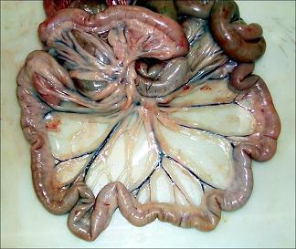
The small intestine extends from the pylorus of the stomach to the caecum . It is attached along it's whole length to the dorsal abdominal wall by mesentry. The mesentry is relatively long for its most part, giving the small intestine a great deal of mobility. The small intestine produces enzymes for digestion of protein, carbohydrate and fat and absorbs the products of their digestion. Enzymes are produced by glands in the intestinal wall and the pancreas. The gall bladder produces bile which emulsifies fats for digestion. Absorption is facilitated by ridges in the small intestine and by the presence of villi and microvilli.
The small intestine consists of three parts:
- duodenum
- jejunum
- ileum.
Duodenum
- Proximal part of the small intestine.
- It has descending and ascending portions.
- The descending duodenum passes out of the pylorus of the stomach (on the right side of the abdomen) and has a sigmoid flexure. It passes towards the right abdominal wall and rises dorsally. In its passage it is related dorsally to the right lobe of the pancreas, ventrally to the jejunum and medially to the ascending colon and caecum.
- At a point between the right kidney and pelvic inlet it turns medially and cranially around the root of the mesentry to become the ascending duodenum. The point of turn is called the caudal flexure of the duodenum.
- The ascending duodenum is shorter and bends ventrally to enter the mesentery and becomes the jejunum.
- Both the pancreatic and the bile duct open into the duodenum.
- Mesoduodenum attaches the duodenum to the dorsal abdominal wall. This is relatively short in the horse and ruminant and longer in the carnivore and pig.
Jejunum
- The jejunum is the longest part of the small intestine.
- It is highly coiled and occupies the ventral part of the abdominal cavity, filling those parts that are not occupied by other viscera. This produces species variation (see comparative aspects).
- It is suspended by the mesentry (mesojejunum). This conveys the blood vessels and nerves and houses lymph nodes.
- The mesentry converges to its root. This is where the cranial mesenteric artery branches off from the aorta.
Ileum
- The ileum is the terminal portion of the small intestine.
- The boundary between the ileum and jejunum is arbitrarily distinguished by the position of the ileocaecal fold.
- It is more muscular and firmer than the jejunum.
- It terminates at the ileocaecocolic junction.
Histology
The intestinal wall is composed of four layers (from inside to outside):
- mucosa
- epithelium
- lamina propria
- lamina muscularis
- submucosa
- tunica muscularis
- circular muscle layer
- longitudinal muscle layer
- serosa
Duodenum
- The mucosa is arranged into villi that provide a large surface area for absorption.
- Epithelium is simple columnar - ideal for absorption.
- Epithelial cells are known as enterocytes.
- A single layer of enterocytes overlies the lamina propria.
- Enterocytes originate from progenitor cells that migrate from mucosal crypts. They differentiate as they migrate up the villus.
- Enterocytes ane absorptive and posses microvilli.
- Membrane bound enzymes and transport proteins are also within the epithelium.
- Each villus houses capillaries that transport amino acids, monosaccharides and other digestive products and lacteals that transport triacylglycerides. Lacteals drain into the lymphatic system.
- Crypts are present at the base of each villus in the mucosa. Cell types in mucosal crypts (from luminal to basal):
- goblet at the tip of the crypt. Produce mucous by exocytosis.
- entero-endocrine in the middle of the crypt. Secrete peptides and amines.
- paneth at the base of the crypt. Function unknown. Contain eosinophilic granules.
- Brunner's glands are present in the submucosa.
- They produce an alkaline secretion which neutralises stomach acid.
- They open into the crypts above in the mucosa.
Jejunum
Ileum
- Peyer's Patches
Comparative Aspects
asd
