Difference between revisions of "Sublingual Gland - Anatomy & Physiology"
Jump to navigation
Jump to search
| Line 12: | Line 12: | ||
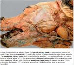
[[Image:Salivary Glands Dog.jpg|thumb|right|150px|Salivary Glands Labelled (Dog) - Copyright C.Clarkson and T.F.Fletcher University of Minnesota]] | [[Image:Salivary Glands Dog.jpg|thumb|right|150px|Salivary Glands Labelled (Dog) - Copyright C.Clarkson and T.F.Fletcher University of Minnesota]] | ||
==[[Sublingual Gland Histology|Histology]]== | ==[[Sublingual Gland Histology|Histology]]== | ||
| − | |||
| − | |||
==Species Differences== | ==Species Differences== | ||
Revision as of 16:36, 4 September 2010
Sublingual Salivary Gland
- Merocrine secretion
- Smaller than parotid gland
- Consists of monostomatic compact part drained by a single duct. Located over rostral mandibluar gland in dog. Duct runs with mandibluar gland duct and opens at the sublingual caruncle.
- Consists of polystomatic diffuse part drained by numerous smaller ducts. Thin strip below mucosa of the floor of oral cavity and duct opens beside the frenulum.
Histology
Species Differences
Ruminant and Pig
Equine
- Diffuse part is the only part of the sublingual salivary gland present
