Difference between revisions of "Alimentary Case 1 Ultrasound"
Jump to navigation
Jump to search
| Line 2: | Line 2: | ||
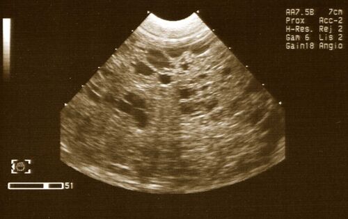
[[Image:Alimentary Case 1 Ultrasound.jpg|thumb|center|500px|Ultrasound Image (Courtesy of A. Antonczyk)]] | [[Image:Alimentary Case 1 Ultrasound.jpg|thumb|center|500px|Ultrasound Image (Courtesy of A. Antonczyk)]] | ||
| + | |||
| + | In order to reveal an answer, highlight the underlined or bulleted area using your mouse. | ||
| + | The number of bullet points doesn't necessarily indicate a strict number of answers. | ||
| + | |||
| + | <big>'''Vet's thoughts'''</big> | ||
| + | |||
| + | *What kind of an image is this? Ultrasound | ||
*Does the image of liver look normal? | *Does the image of liver look normal? | ||
| + | **No | ||
| + | **Scan of the liver made a tumour affecting the liver very likely | ||
| − | * | + | *Other points: |
| − | + | **The ultrasound confirmed presence of fluid within the abdomen (image not shown) | |
| − | |||
| − | |||
| − | |||
| − | |||
Revision as of 14:32, 3 December 2007
In order to reveal an answer, highlight the underlined or bulleted area using your mouse.
The number of bullet points doesn't necessarily indicate a strict number of answers.
Vet's thoughts
- What kind of an image is this? Ultrasound
- Does the image of liver look normal?
- No
- Scan of the liver made a tumour affecting the liver very likely
- Other points:
- The ultrasound confirmed presence of fluid within the abdomen (image not shown)
