Difference between revisions of "Toxocara canis"
| Line 33: | Line 33: | ||
====Cycle 3==== | ====Cycle 3==== | ||
| − | In the pregnant bitch larvae that have become hypobiotic as described in cycle 2 above are reactivated by hormonal changes. These larvae become mobile about three weeks before parturition and migrate across the placenta to the lungs of the fetus. Within the fetal lungs the larvae moult just prior to birth. From the lungs the larvae complete their life cycle in the same way as in the young animal, by being coughed and swallowed to enter the small intestine. The adults will then produce eggs which are released in the faeces as normal. | + | In the pregnant bitch larvae that have become hypobiotic as described in cycle 2 above are reactivated by hormonal changes. These larvae become mobile about three weeks before parturition and migrate across the placenta to the lungs of the fetus. Within the fetal lungs the larvae moult just prior to birth (to L3). From the lungs the larvae complete their life cycle in the same way as in the young animal, by being coughed and swallowed to enter the small intestine. The adults will then produce eggs which are released in the faeces as normal. |
| + | |||
====Cycle 4==== | ====Cycle 4==== | ||
The final life cycle involves transmission of L3 larvae to pups through the milk. Hypobiotic L3 larvae are reactivated and are either already present in the mammary glands or travel to them and are capable of passing in the milk during the first 3 weeks of lactation. There is no further migration in the pup when the larvae are ingested in this way and the remaining life cycle of the worm is completed in the small intestine of the pup. | The final life cycle involves transmission of L3 larvae to pups through the milk. Hypobiotic L3 larvae are reactivated and are either already present in the mammary glands or travel to them and are capable of passing in the milk during the first 3 weeks of lactation. There is no further migration in the pup when the larvae are ingested in this way and the remaining life cycle of the worm is completed in the small intestine of the pup. | ||
Revision as of 21:28, 28 April 2013
| This article is still under construction. |
| Toxocara canis | |
|---|---|
| Kingdom | Animalia |
| Phylum | Nematoda |
| Class | Secernentea |
| Order | Ascaridida |
| Family | Toxocaridae |
| Genus | Toxocara |
| Species | T. canis |
Overview
Toxacara canis is a typical ascarid nematode that infects dogs where its predilection site is the small intestines. These worms can be found throughout the world with varying prevalence. Control of this ascarid is typically difficult due to its extended persistence in the environment. T. canis is also important in human medicine as the species most responsible for visceral larval migrans (VLM). The human is a non-permissive host of T. canis meaning it cannot complete its life cycle and reproduce, however the larval stages do migrate through the human body causing pathology.
Identification
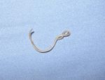
Toxacara canis has the typical gross morphology of an ascarid, it is a large, fleshy white worm and can be up to 18cm long. The females are longer than the males who can normally reach 10cm in length. Microscopically T. canis has a fairly standard ascaridoid appearance, though the adult head is given an elliptical shape by large alae or 'wings'. The eggs of T. canis are dark brown with a thick, pitted shell, the thick shell makes them very resistant in the environment.
Life cycle
Typically of an ascarid T. canis has larvae have a migratory life cycle that is significance in the pathogenesis of infection. This species also has the most complex life cycle in the Ascaridoidea superfamily. There are four different life cycles that can occur dependant on the circumstances that the larvae or adult encounter.
Cycle 1
This is mostly a typical ascarid life cycle and commonly occurs in dogs that are infected between 2 and 3 months old. The infective eggs contain L2 larvae which hatch in the small intestine of the host dog after being ingested. The larvae then enter the hepatic portal vein and travel through the liver and further to the lungs where they moult to L3. The larvae then migrate to the trachea where they are coughed up and swallowed again by the host. This is known as hepato-tracheal migration. On returning to the small intestine they undergo the final moults (L3-->L4-->L5) before becoming adults.
Cycle 2
In older dogs (above 3 months) the migration changes and the hepato-tracheal route occurs far less often, though can still occur. In these animals the L2 larvae hatch in the small intestine and travel to a wide variety of tissues throughout the body. Once the larvae have reached a tissue they will begin hypobiosis and encyst in the tissue until reactivated. In some animals the hypobiotic larvae will not reactivate and this will be the end of their life cycle. Hypobiotic larvae in the tissues of the dog are known as somatic larvae, although these do not grow or develop they are highly metabolically active. The produce large quantities of excretory/secretory antigens which are spread over the cuticle of the worm. These antigens are important in immune evasion by way of having a rapid turnover and sloughing off host antibodies and immune cells.
Cycle 3
In the pregnant bitch larvae that have become hypobiotic as described in cycle 2 above are reactivated by hormonal changes. These larvae become mobile about three weeks before parturition and migrate across the placenta to the lungs of the fetus. Within the fetal lungs the larvae moult just prior to birth (to L3). From the lungs the larvae complete their life cycle in the same way as in the young animal, by being coughed and swallowed to enter the small intestine. The adults will then produce eggs which are released in the faeces as normal.
Cycle 4
The final life cycle involves transmission of L3 larvae to pups through the milk. Hypobiotic L3 larvae are reactivated and are either already present in the mammary glands or travel to them and are capable of passing in the milk during the first 3 weeks of lactation. There is no further migration in the pup when the larvae are ingested in this way and the remaining life cycle of the worm is completed in the small intestine of the pup.
As well as the above life cycles T. canis can infect paratenic hosts such as mice, rats and some birds. Events occur just as in the older dog, i.e. larvae migrate → liver → lungs → heart → somatic tissues → granulomatous reactions → 'waiting phase'; but in this case, the somatic larvae are waiting for the animal that they are in (which is acting as a paratenic host) to be eaten by a dog, fox, wolf or other canid, where they will establish as adults or somatic larvae (depending on the age of the predator). This explains how humans (as warm-blooded non-canid animals) enter into the epidemiological picture. The prepatent period of T. canis is 4 - 5 weeks in the canid host.
Epidemiology
T. canis is present worldwide with a wide range of prevalances in different areas from 5 - 80%. Adult animals carry the fewest worms since initial infection causes immunity which leads to the shedding of adult worms from the intestines. The low parasite burden in adult animals can often lead to asymptomatic infection though the parasites wil still shed eggs in the faeces. The largest numbers of worms are found in dogs less than 6 months old who have not yet gained immunity to the worms. The high levels of prevalence of this species worldwide is largely due to the difficulty in controlling its spread. The eggs are extremely resistant in the environment and so can persist for several years. The females lay very large numbers of eggs, up to 700 per gram of faeces, making the removal of such a large number difficult. This final reason for such a large spread is the long lasting reservoir of hypobiotic larvae that can be reactivated in pregnancy in the bitch, these are not susceptible to anthelmintics and so are only eliminated by preventing pregnancy or the death of the host. As a result of the large number of infected bitches almost all puppies are born with T. canis infections which increases the spread of the eggs as they pass faeces in new environments once the litter is split up.
Pathogenesis
In puppies heavy infections can cause weight loss and poor growth as well as diarrhoea and vomiting. Pot belly may also be seen in pupies in some cases. With extremely heavy infections a plug may form that can cause intestinal impaction and prevent gastric movements. In adults, once immunity has developed, there are few clinical signs as most infections are too small for pathology to develop. In humans there is a zoonotic risk, as T. canis is the major agent of visceral larval migrans in children primarily with occular migration.
Control
Control of T. canis relies on effective clearing of the eggs form the environment as these can be infective in the environment for several years. This will prevent new infections of animals that have no been exposed previously as pups or as young dogs. However there are a number of endemic regions of the world where most animals have been exposed as pups and therefore can harbour hypobiotic larvae. These are difficult to eliminate and there are likely to be constantly be a small number of worms present, therefore regular treatment of dogs with anthelmintics is recommended.
- Causes eosinophilic enteritis in the dog
In warm-blooded non-canid animals:
- Events occur just as in the older dog, i.e. larvae migrate → liver → lungs → heart → somatic tissues → granulomatous reactions → 'waiting phase'; but in this case, the somatic larvae are waiting for the animal that they are in (which is acting as a paratenic host) to be eaten by a dog, fox, wolf or other canid, where they will establish as adults or somatic larvae (depending on the age of the predator).
- This explains how humans (as warm-blooded non-canid animals) enter into the epidemiological picture.
Epidemiology
- Infection of dogs is by ingestion of the L2 larvae, which can occur in four ways:
1) ingestion of the embryonated egg
2) prenatal infection
3) transmammary infection
4) ingestion of a paratenic host.
- Infection of a paratenic host can occur by:
1) ingestion of the embryonated egg
2) ingestion of larvae in the tissues of another paratenic host.
- Each female T. canis can lay up to 250,000 eggs per day:
- the eggs are not infective until the L2 is fully developed
- this process takes a few weeks in summer, but many weeks in the winter
- the embryonated egg is tough and can survive for 4-5years
- eggs therefore accumulate in the environment, and can easily be demonstrated in soil scrapings from, for example, breeding kennels or city parks
- when eggs from the environment are swallowed by a bitch, larvae accumulate in her somatic tissues - to be activated during pregnancy
- at birth, prenatally derived larvae are already migrating through the pups' liver and lungs
- adult worms reach the intestine and start to lay eggs when the pups are 2-3weeks old
- pups are therefore a potent source of environmental contamination (particularly in breeding kennels) until spontaneous expulsion occurs after approximately 6weeks of age.
- in general, only approximately 15% of adult dogs have patent infection - an exception is nursing bitches, who often pass large numbers of eggs
- up to 45% of foxes have patent infection, and are therefore a potent source of eggs in urban areas.
Human Infection
- Humans are infected by swallowing embryonated eggs from the environmental reservoir.
- This is most likely to happen in young children.
- Most infections are asymptomatic.
- Approsimately 2.5% of the British population are seropositive.
Efficacy of Anthelmintics Against Life-Cycle Stages of T. canis
| Compound | Trade-Name | Intestinal Worms | Migrating Larvae | Somatic Larvae |
|---|---|---|---|---|
|
Piperazine
|
various
|
+
|
-
|
-
|
Control of T. canis
- The only satisfactory way of breaking the life-cycle in breeding kennels and reducing zoonotic risk is to eliminate T. canis eggs from the environment.
- Hygiene is important (but note that the eggs stick to surfaces and that few disinfectants will kill them).
- To prevent dogs excreting eggs, pups must be dosed regularly from 2weeks of age.
- Most anthelmintics are only active against adult worms in the intestine.
- These adult worms are quickly replaced by developing larvae that survived treatment.
- Therefore, pups should be dosed at 2, 4, 6, 8 and 12weeks of age.
- Fenbendazole is active against both adults and larvae.
- So, an equivalent result can be obtained with just two treatments: one in the third week of life, and again 3weeks later.
- Nursing bitches should also be treated.
- Otherwise, adult dogs should be dosed 2-4times a year.
- Current anthelmintics at normal dose-rates will not kill somatic larvae.
- This can be done, however, with daily high doses of fenbendazole.
- Pregnant bitches are given daily doses (25mg/kg) from the 42nd day of pregnancy.
T. canis in Veterinary Public Health
T. canis is associated with at least three disease syndromes in humans:
1) visceral larval migrans (VLM) (→ eosinophilia, hepatomegaly, fever, asthma)
2) ocular larval migrans (OLM) (→ unilateral partial impairment of vision)
3) covert toxocarosis (non-specific clinical signs associated with high antibody titre)
Around 55cases, mostly OLM, are diagnosed in the UK each year.

