Difference between revisions of "Muscles - Anatomy & Physiology"
Jump to navigation
Jump to search
| Line 9: | Line 9: | ||
==Skeletal Muscle== | ==Skeletal Muscle== | ||
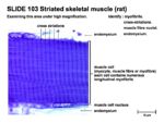
| + | [[Image:Striated Muscle 1.jpg|thumb|right|150px|Striated muscle - Copyright RVC 2008]] | ||
*Each muscle is composed of multiple '''fascicles''', each of which consists of a bundle of muscle fibers | *Each muscle is composed of multiple '''fascicles''', each of which consists of a bundle of muscle fibers | ||
*Muscle "fiber" means a single cell, which are multi-nucleate, and known as '''syncitia''' | *Muscle "fiber" means a single cell, which are multi-nucleate, and known as '''syncitia''' | ||
Revision as of 09:19, 23 July 2008
Introduction
Muscle mass accounts for a large majority of the carcass weight of domestic animals. Muscular contraction is necessary for voluntary and involuntary movement of limbs, stabilization of joints, maintaining luminal diameter (in the case of arteries, bowel, etc), and to produce heat. The number of muscle fibers an individual possesses is determined by genetics and is set at birth, although muscle size and type (i.e. glycolytic to oxidative and vice versa) can be altered. Further muscle development therefore occurs by hypertrophy, rather than hyperplasia of muscle fibers. Three types of muscle can be described:
- Skeletal (aka Striated, Somatic, Voluntary)
- Smooth (aka Visceral)
- Cardiac
Skeletal Muscle
- Each muscle is composed of multiple fascicles, each of which consists of a bundle of muscle fibers
- Muscle "fiber" means a single cell, which are multi-nucleate, and known as syncitia
- Within each fiber are groups of parallel, longitudinal myofibrils
- Myofibrils are arranged as sarcomeres, bound by Z-discs, which are the functional unit of muscle contraction
- Each sarcomere contains 2 separate groups of myofilaments:
- Thin filament, containing Actin, located centrally
- Thick filament, containing Myosin, originating from either side of each Z-disc
- Two basic types of skeletal myofibre:
- Primary: Oxidative
- Grossly red
- High myoglobin level
- Slow rate of contraction
- High oxidative activity
- Function - postural, sustained activity
- Secondary: Glycolytic
- Grossly white
- Low myoglobin level
- Fast rate of contraction
- High glycolytic activity
- Function - exercise, bursts of activity
- Primary: Oxidative
- Muscle activation is initiated by a nervous impulse crossing the Neuromuscular Junction
- The neurotransmitter, Acetylcholine (Ach), binds receptors in the muscle fiber to open Na+ channels
- This causes a wave of depolarization along the sarcoplasmic membrane, further opening voltage-gated Na+ channels, which propagates the signal along the sarcolemma
- Depolarization of the sarcoplasmic reticulum causes Calcium to be released, which activates muscle contraction
- Muscle contraction occurs when (thin) Actin filaments slide past (thick) Myosin filaments
- Myosin heads bind Actin subunits, forming cross-bridges, hydrolyzing ATP and providing energy for contraction
- Myosin heads undergo power stroke, displacing Actin and releasing ADP and Pi
- Calcium regulates muscle contraction by binding troponin-C, which is attached to the thin filament
- This causes inhibition of the steric block keeping Actin and Myosin from interacting
- Increased Calcium causes a negative feedback inhibition of Ca release, and it is pumped back into the sarcoplasmic reticulum by the Ca/ATPase pump
Tendon
- Consists of dense collagen type 1 fibres and fibroblasts (tenocytes)

