Difference between revisions of "Cutaneous Larva Migrans"
JamesSwann (talk | contribs) |
JamesSwann (talk | contribs) |
||
| Line 2: | Line 2: | ||
==Description== | ==Description== | ||
| − | [[File:Larva Migrans Cutanea.jpg|thumb|Image of a human foot affected by CLM<br><small>Copyright WeisSagung 2009 Wikimedia Commons]] | + | [[File:Larva Migrans Cutanea.jpg|thumb|Image of a human foot affected by CLM<br><small>Copyright WeisSagung 2009 Wikimedia Commons]]</small> |
| − | Cutaneous Larva Migrans (CLM) refers to a cutaneous reaction to the migration of zoonotic parasite larvae within the skin. The major causes of CLM in humans in the UK are the larvae of the [[:Category:Ancylostomatoidea|hookworms]], [[ | + | Cutaneous Larva Migrans (CLM) refers to a cutaneous reaction to the migration of zoonotic parasite larvae within the skin. The major causes of CLM in humans in the UK are the larvae of the [[:Category:Ancylostomatoidea|hookworms]], ''[[Ancylostoma caninum]]'' and ''[[Uncinaria stenocephala]]''. Other causative larvae are those of the avian schistosomes, ''Strongyloides westeri'' and ''papillosus'' and ''Pelodera strongyloides''. |
| − | In CLM caused by hookworms, the L3 larvae penetrate the skin and migrate for up to two weeks before they are killed by the development of an immune response. In areas where hookworms are endemic, the larvae are acquired where bare skin comes into contact with sand or warm moist soil. The larvae are thought to lack the collagenolytic enzymes that would allow them to penetrate into the dermis and complete their life-cycle and instead, they continue to migrate in the superficial layers of the skin at a rate of up to 2 cm per day. Small serpiginous (snake-like) tunnels may be seen to radiate from the initial point of penetration. The disease is usually treated with thiabendazole. | + | In CLM caused by hookworms, the L3 larvae penetrate the skin and migrate for up to two weeks before they are killed by the development of an immune response. In areas where hookworms are endemic, the larvae are acquired where bare skin comes into contact with sand or warm moist soil. The larvae are thought to lack the collagenolytic enzymes that would allow them to penetrate into the dermis and complete their life-cycle and instead, they continue to migrate in the superficial layers of the skin at a rate of up to 2 cm per day. |
| + | |||
| + | ==Diagnosis== | ||
| + | The affected person will have a history of exposure to the larvae of the hookworms, either from travel to an endemic area or if they are exposed to larvae derived from dogs in the UK. | ||
| + | |||
| + | ===Clinical Signs=== | ||
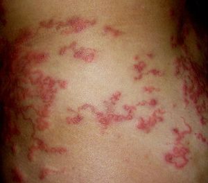
| + | Small serpiginous (snake-like) tunnels may be seen to radiate from the initial point of penetration, as seen in the image. The area around these tunnels is inflamed and may be intensely pruritic. | ||
| + | |||
| + | ==Treatment== | ||
| + | The disease is usually treated with thiabendazole. | ||
[[Category:Zoonoses]][[Category:To_Do_-_James]] | [[Category:Zoonoses]][[Category:To_Do_-_James]] | ||
Revision as of 10:08, 20 July 2010
| This article is still under construction. |
Description
Cutaneous Larva Migrans (CLM) refers to a cutaneous reaction to the migration of zoonotic parasite larvae within the skin. The major causes of CLM in humans in the UK are the larvae of the hookworms, Ancylostoma caninum and Uncinaria stenocephala. Other causative larvae are those of the avian schistosomes, Strongyloides westeri and papillosus and Pelodera strongyloides.
In CLM caused by hookworms, the L3 larvae penetrate the skin and migrate for up to two weeks before they are killed by the development of an immune response. In areas where hookworms are endemic, the larvae are acquired where bare skin comes into contact with sand or warm moist soil. The larvae are thought to lack the collagenolytic enzymes that would allow them to penetrate into the dermis and complete their life-cycle and instead, they continue to migrate in the superficial layers of the skin at a rate of up to 2 cm per day.
Diagnosis
The affected person will have a history of exposure to the larvae of the hookworms, either from travel to an endemic area or if they are exposed to larvae derived from dogs in the UK.
Clinical Signs
Small serpiginous (snake-like) tunnels may be seen to radiate from the initial point of penetration, as seen in the image. The area around these tunnels is inflamed and may be intensely pruritic.
Treatment
The disease is usually treated with thiabendazole.
