Difference between revisions of "File:Ultrasound testis donkey.jpg"
Jump to navigation
Jump to search
(Courtesy of The Donkey Sanctuary) |
|||
| Line 1: | Line 1: | ||
| − | + | {{Information | |
| + | |Description=Ultrasonography of the testis and epididymis. Longitudinal view of the testis showing testicular parenchyma (T) and the central vein of the mediastinum testis (arrow). | ||
| + | |Source=The Donkey Sanctuary | ||
| + | |Date=unknown | ||
| + | |Author=unknown | ||
| + | |Permission=See below | ||
| + | |Other_versions= | ||
| + | }} | ||
| + | |||
| + | {{cc-att-2.0}} | ||
Latest revision as of 10:29, 25 October 2010
| Description |
Ultrasonography of the testis and epididymis. Longitudinal view of the testis showing testicular parenchyma (T) and the central vein of the mediastinum testis (arrow). |
|---|---|
| Date |
unknown |
| Source |
The Donkey Sanctuary |
| Author |
unknown |
| Permission (Reusing this file) |
See below |
| This file is licensed under the Creative Commons Attribution Non-Commercial & No Derivative Works License |
File history
Click on a date/time to view the file as it appeared at that time.
| Date/Time | Thumbnail | Dimensions | User | Comment | |
|---|---|---|---|---|---|
| current | 13:15, 3 February 2010 | 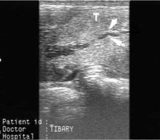 | 536 × 470 (21 KB) | Bara (talk | contribs) | Courtesy of The Donkey Sanctuary |
You cannot overwrite this file.
File usage
The following 2 pages use this file: