Small Animal Soft Tissue Surgery Q&A 19
| This question was provided by Manson Publishing as part of the OVAL Project. See more Small Animal Soft Tissue Surgery Q&A. |
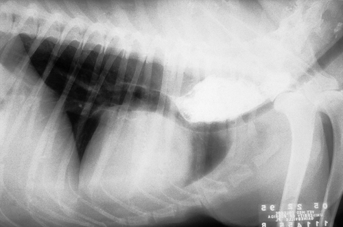
You are presented with a 12-week-old, male Weimaraner puppy with a history of regurgitation noticed shortly after the puppy was purchased from the breeder. On physical examination the puppy is thin but alert and active. The owners report that he does not regurgitate liquids. Survey radiographs of the chest and abdomen are normal. The barium swallow results are shown.
| Question | Answer | Article | |
| What is the most likely diagnosis? | Vascular ring anomaly. Persistent right aortic arch is most likely. A breed predisposition is identified for Weimaraners, Great Danes, Irish Setters and Doberman Pinschers. |
Link to Article | |
| Describe the pathophysiology of this condition. | Persistent right aortic arch results from failure of the left aortic arch to develop as the dominant structure in the fetus; instead, the right aortic arch develops. Cardiovascular physiology is normal but the ligamentum arteriosum (ductus arteriosus of the fetus) connects the right-sided aorta to the pulmonary artery creating the vascular ring entrapping the esophagus and trachea. The primary clinical sign is regurgitation at weaning. Emaciation and aspiration pneumonia may occur secondarily. |
Link to Article | |
| How should this dog be managed prior to surgery, and when should surgery be performed? | Surgery is performed as soon as possible. If aspiration pneumonia is present, it is treated first. If surgery is delayed, the esophagus may develop permanent loss of muscle tone and neurologic dysfunction. Prior to surgery, the puppy is fed a liquid or gruel diet (small amounts frequently, from an elevated position) which will pass through the vascular ring. |
Link to Article | |
| Describe the surgical management of this condition. | Surgery involves transection of the ligamentum arteriosum and freeing the esophagus from its entrapment within the vascular ring. The ligamentum arteriosum, approached via left lateral 4th intercostal thoracotomy, is identified caudal to the esophageal dilation and dissected free from attachments to the esophagus. Because it can be patent, the ligament is double ligated and transected between ligatures. A large orogastric tube is inserted into the esophagus and passed through the constricted area; any tissue preventing distension is dissected free allowing the tube to pass easily. |
Link to Article | |
| What is the long-term prognosis for this dog, and how should it be managed postoperatively? | Prognosis with early surgical treatment is good. The longer surgery is delayed, the poorer the prognosis. Postoperative management is identical to preoperative management. Four weeks following surgery the puppy is gradually returned to a normal diet. If regurgitation occurs, a barium swallow is repeated to determine the status of the esophagus. |
Link to Article | |
