File:VSD1.jpg
Jump to navigation
Jump to search

Size of this preview: 800 × 546 pixels. Other resolutions: 320 × 218 pixels | 1,157 × 790 pixels.
Original file (1,157 × 790 pixels, file size: 90 KB, MIME type: image/jpeg)
Summary
| Description |
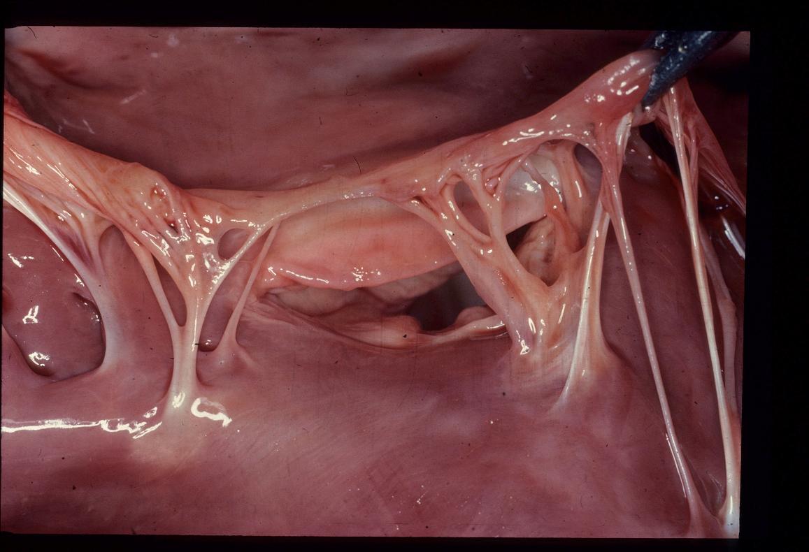
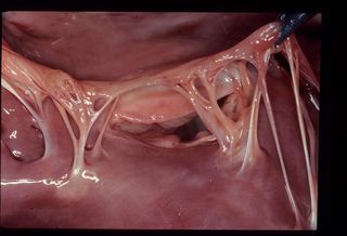
Ventricular septal defects are most commonly seen high in the interventricular septum, in the membranous region, as seen with this defect. The defect is just underneath the AV valves and allows left to right blood flow, overloading the right ventricle. |
|---|---|
| Date |
Unknown |
| Source |
Cambridge University |
| Author |
A. Jefferies |
| Permission (Reusing this file) |
See below |
Licensing:
| This file is licensed under the Creative Commons Attribution Non-Commercial & No Derivative Works License |
File history
Click on a date/time to view the file as it appeared at that time.
| Date/Time | Thumbnail | Dimensions | User | Comment | |
|---|---|---|---|---|---|
| current | 15:32, 28 August 2007 | | 1,157 × 790 (90 KB) | Kjr35 (talk | contribs) |
You cannot overwrite this file.
File usage
The following 2 pages use this file: