File:Pericarditis-histo.jpg
Jump to navigation
Jump to search

Size of this preview: 800 × 546 pixels. Other resolutions: 320 × 218 pixels | 1,157 × 790 pixels.
Original file (1,157 × 790 pixels, file size: 160 KB, MIME type: image/jpeg)
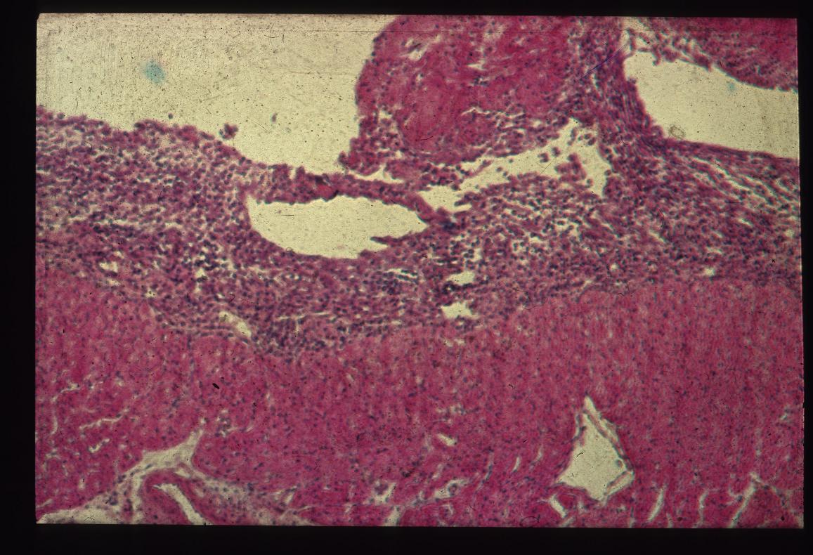
The epicardium should consist of a single layer of epithelial cells in the normal heart with a similar layer lining the opposing pericardial surface. In this histological section there is an increase in cellularity of the epicardium with many inflammatory cells and pink fibrin is visible.
File history
Click on a date/time to view the file as it appeared at that time.
| Date/Time | Thumbnail | Dimensions | User | Comment | |
|---|---|---|---|---|---|
| current | 15:41, 29 August 2007 | | 1,157 × 790 (160 KB) | Kjr35 (talk | contribs) |
You cannot overwrite this file.
File usage
The following 2 pages use this file: