File:Perianal gland hepatoid adenoma dog.jpg
Jump to navigation
Jump to search
Perianal_gland_hepatoid_adenoma_dog.jpg (478 × 277 pixels, file size: 37 KB, MIME type: image/jpeg)
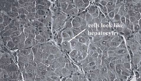
Perianal gland "hepatoid" adenoma in the dog (benign). Note the well differentiated cells, that resemble hepatocytes. The tissue is well organised, there are few mitoses and there is no haemoorhage or necrosis.
Pending permission from Brian Smyth.
File history
Click on a date/time to view the file as it appeared at that time.
| Date/Time | Thumbnail | Dimensions | User | Comment | |
|---|---|---|---|---|---|
| current | 11:17, 3 September 2007 |  | 478 × 277 (37 KB) | Lizzies (talk | contribs) | Perianal gland "hepatoid" adenoma in the dog (benign). Note the well differentiated cells, that resemble hepatocytes. The tissue is well organised, there are few mitoses and there is no haemoorhage or necrosis. Pending permission from Brian Smyth. |
You cannot overwrite this file.
File usage
There are no pages that use this file.
