| Strongyloides spp. | |
|---|---|
| Kingdom | Animalia |
| Phylum | Nematoda |
| Class | Chromadorea |
| Order | Rhabditida |
| Sub-order | Rhabditina |
| Super-family | Rhabditoidea |
| Family | Strongyloididae |
| Genus | Strongloides |
| Species | S. westeri |
Overview
This is the only genus of veterinary importance in the super-family Rhabditoidea. There are a number of species that are of particular importance and these are mainly host specific.
Strongyloides westeri
This is a parasite of the horse and is most commonly seen in young foals, less than six months in age. This is usually the fisrt parasite to which foals are exposed and therfore they have very little immunity to these infections. The predeliction site for these worms is the small intestine.
Morphology
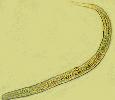
The distinguishing features of this worm are:
- Very small, 6-9mm long
- Slender, hair-like
- Long oesophagus (up to one third of body length).
- Intestines and ovaries intertwined in caudal body
- Blunt ended tail
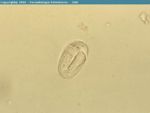
The eggs are small and oval with a thin shell, they will often hatch in the large intestine so L1 larvae are expelled in the hosts faeces.
Life-Cycle
S. westeri has a typical strongyloides life cycle, with both a free living and parasitic cycle. The free living cycle involves male and female worms reproducing sexually on the ground, this can occur for several generation with no parasitism taking place. The parasitic phase involves only female worms and occurs from ingesion of L£ larvae or their penetration through the hosts skin. Infection my also be transmissed vertically from the dam to the foal by larval migration to the mammary tissue and ingestion by the foal with the dams milk. Once in the host the larvae migrate to the small intestine where they tunnel into the epithelium at the base of the villi and moult to L4 and then adult females. Females produce eggs without sexual interaction that can develop to become both male and female larvae. The pre patent period is between 1 and 2 weeks dependant on the level of infection.
Pathogenicity
Pathology is rarely seen in adult horses due to the development of immunity, though penetration of the skin by larvae may still cause irritation anddermatitis. Adults with larvae in beneath the skin and in the abdomen can act as carriers. In foals heavy infections can cause severe enteritis and diarrhoea. It should be noted that apparently healthy animals can still have high faecal egg counts.
Control
The number and spread of S. westeri is best controlled by good hygeine such as the removal of faeces from pasture and stabling and the provision of clean, dry bedding. Foals are often treated with anthelmintics at 2 weeks old against S. westeri.
Strongyloides papillosus
This is a nematode of cattle and small ruminants and is found throughout the world. As with ‘’S. westeri’’ above clinical signs are normally only seen in young animals as older animals develop immunity.
Morphology
The morphology of S. papillosus’’ is the same as that of the ‘’S. westeri’’. The eggs hatch inside the host and so L1 larvae are released with the faeces.
Life Cycle
The life cylce of S. papillosus is typical of strongyloides species with both parasitic and free living cycles. As adults animals will become immune to infection the primary method of infection for young animals is through the milk of the dam, this still involves only females as males are only present in the environment as 'free-living' nematodes.


