File:Equine Internal Medicine Q&A 11B.jpg
Revision as of 14:50, 31 May 2011 by Ggaitskell (talk | contribs) ({{Information |Description=Image from [http://www.mansonpublishing.com/vet_titles/Meir.html 'Equine Internal Medicine'], with permission from Manson Publishing, as part of the OVAL Project. This is an ultrasound of a foal with a urachal di)
Equine_Internal_Medicine_Q&A_11B.jpg (500 × 348 pixels, file size: 151 KB, MIME type: image/jpeg)
Summary
| Description |
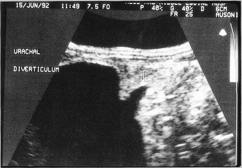
Image from 'Equine Internal Medicine', with permission from Manson Publishing, as part of the OVAL Project. This is an ultrasound of a foal with a urachal diverticulum. |
|---|---|
| Date |
uploaded to WikiVet 31/05/2011 |
| Source | |
| Author |
Manson Publishing |
| Permission (Reusing this file) |
See below |
Licensing
| This file is licensed under the Creative Commons Attribution Non-Commercial & No Derivative Works License |
File history
Click on a date/time to view the file as it appeared at that time.
| Date/Time | Thumbnail | Dimensions | User | Comment | |
|---|---|---|---|---|---|
| current | 14:50, 31 May 2011 |  | 500 × 348 (151 KB) | Ggaitskell (talk | contribs) | {{Information |Description=Image from [http://www.mansonpublishing.com/vet_titles/Meir.html 'Equine Internal Medicine'], with permission from Manson Publishing, as part of the OVAL Project. This is an ultrasound of a foal with a urachal di |
You cannot overwrite this file.
File usage
The following 2 pages use this file:
