|
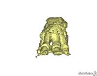
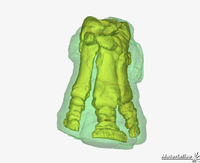
3D imaging of the bones in elephant and rhino by John Hutchinson, used to teach 3D skeletal anatomy.
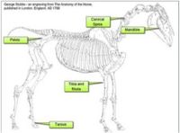
Interactive drag and drop activity based on Stubbs work from 1766. Accurate and detailed drawings based on dissection showing the full anatomy of the horse used to create interactive exercises for revision and learning.
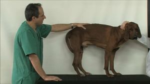
Equine locomotion by Renate Weller. Showing a horse trotting in slow motion focusing on the lower leg and the movement of joints through realtime radiography.
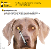
Interactive resource testing the functional integrity of the ocular reflex on a virtual patient in the form of a Flash animation. An example of a resource which helps to integrate anatomy teaching with the clinical years. Recently awarded first prize at JORUM, a national award for the best OER.
| 