- Neurons are particularly vulnerable to injury, due to their:
- High metabolic rate
- Small capacity to store energy
- Lack of regenerative ability
- Axons being very dependent on the cell body.
- Axons cannot make their own protein as they have no Nissl substance.
- The cell body produces the axon's protein and disposes of its waste.
- Death or damage of the cell body causes axon degeneration.
- There are four ways in which neurons may react to insult:
- Acute Necrosis
- Chromatolysis
- Wallerian Degeneration
- Vacuolation
Acute Necrosis
- Acute necrosis is the most common neuronal response to injury.
- Causes of actue necrosis include:
- Ischaemia
- Diminution of the blood supply causes a lack of nutrients and oxygen, inhibiting energy production. A decrease in the levels of ATP leads to:
- Failure of the Na+/K+pumps, causing cell swelling and an increase in extracellular potassium.
- Failure to generate NAD required for DNA repair.
- Diminution of the blood supply causes a lack of nutrients and oxygen, inhibiting energy production. A decrease in the levels of ATP leads to:
- Hypoxia
- Hypoglycaemia
- Toxins, such as lead and mercury
- Ischaemia
Laminar Cortical Necrosis
- Laminar cortical necrosis refers to the selective destruction of neurons in the deeper layers of the cerebral cortex.
- These neurons are the most sensitive to hypoxia.
- The laminar cortical pattern of acute necrosis occurs in several instances:
- Ischaemia
- For example, seizure-related ischaemia in dogs.
- Polioencephalomalacia in ruminants
- Also called cerebrocortical necrosis or CCN.
- Salt poisoning in swine
- Lead poisoning in cattle
- Ischaemia
- It is most likely that gross changes will not be seen. When they are visible, changes may be apparent as:
- Oedema
- Causes brain swelling, flattened gyri and herniation
- A thin, white, glistening line along the middle of the cortex.
- In ruminants, this fluoresces with UV-light.
- Oedema
- Ultimately the cortex becomes necrotic and collapses.
Chromatolysis
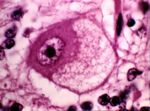
- Chromatolysis is the cell body’s reaction to axonal insult.
- The cell body swells and the Nissl substance (granular cytoplasmic reticulum and ribosomes found in nerve cell bodies) disperses.
- Dispersal of the Nissl substance allows the cell body to produce proteins for rebuilding the axon.
- IT IS NOT A FORM OF NECROSIS.
- It is an adaptive response to deal with the injury.
- It can, however lead to necrosis.
- Seen, for example, in grass sickness in horses (equine dysautonomia).
Wallerian Degeneration
- Wallerian degeneration is the axon’s reaction to insult.
- The axon and its myelin sheath degenerates distal to the point of injury.
- There are several causes of wallerian degeneration:
- Axonal transection
- This is the "classic" cause
- Vascular causes
- Inflamatory reactions
- Toxic insult
- As a sequel to neuronal cell death.
- Axonal transection
View images courtesy of Cornell Veterinary Medicine
The Process of Wallerian Degeneration
- Axonal Degeneration
- Axonal injuries initially lead to acute axonal degeneration.
- The proximal and distal ends separate within 30 minutes of injury.
- Degeneration and swelling of the axolemma eventually leads to formation of bead-like particles.
- After the membrane is degraded, the organelles and cytoskeleton disintegrate.
- Larger axons require longer time for cytoskeleton degradation and thus take a longer time to degenerate.
- Axonal injuries initially lead to acute axonal degeneration.
- Myelin Clearance
- Following axonal degeneration, myelin debris is cleared by phagocytosis.
- Myelin clearance in the PNS is much faster and efficient that in the CNS. This is due to:
- The actions of schwann cells in the PNS.
- Differences in changes in the blood-brain barrier in each system.
- In the PNS, the permeability increases throughout the distal stump.
- Barrier disruption in CNS is limited to the site of injury.
- Regeneration
- Regeneration is rapid in the PNS.
- Schwann cells release growth factors to support regeneration.
- CNS regeneration is much slower, and is almost absent in most species.
- This is due to:
- Slow or absent phagocytosis
- Little or no axonal regeneration, because:
- Oligodendrocytes have little capacity for remyelination compared to Schwann cells.
- There is no basal lamina scaffold to support a new axonal sprout.
- The debris from central myelin inhibits axonal sprouting.
- This is due to:
- Regeneration is rapid in the PNS.
Vacuolation
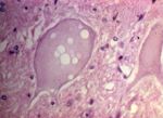
- Vacuolation is the hallmark of transmissible spongiform encephalopathies.
- For example, BSE and Scrapie.
- Vacuolation can also occur under other circumstances:
- Artefact of fixation
- Toxicoses
- It may sometimes be a normal feature.

