| This article is still under construction. |
| Also known as: | Equine lungworm |
Scientific Classification
| Class | Nematoda |
| Superfamily | Trichodectidae |
Hosts
Donkeys, and occasionally horses.
Identification
Adults are slender, thread-like and white. Females are larger than males at around 6.5cm in length. The males have a small non-lobulated bursa.
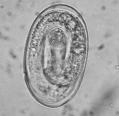
The embryonated eggs are 80-100µm in length.
Life Cycle
The lifecycle is not greatly known, but it is currently thought to be similar to that of Dictyocaulus viviparous.
The prepatent period is 2-3 months.
Epidemiology
- Main source of infection = donkeys (remain infected for years) - contaminate horse pasture.
- Infection can cycle in horses.
| Horses | Donkeys | |
|---|---|---|
| Prevalence | 10-20% | 75% |
| Adult worms | Few | Many |
| Eggs in faeces | Often zero | Many |
| Period of patency | <8months | 5+ years |
| Clinical signs | Sometimes | Rarely |
NOTE: Clinical signs - chronic cough at rest or during exercise, single animal or group of horses, autumn or early winter.
- Dictyocaulus arnfieldi causes cough in horses
Pathogenicity
- Raised areas of over-inflated pulmonary tissue (several cms in diameter) surrounding small bronchus containing worms and mucopurulent exudate.
- Hyperplastic bronchial epithelium.
- Peribronchial "cuffing".
Diagnosis
- Clinical signs.
- Grazing history (donkey contact or shared grazing).
- Faecal examination (only detects patent infections = small proportion of lungworm infections in horses):
- process sample immediately = McMaster method, embryonated eggs
- process sample later = Baerman technique, larvae with tail spine.
- Tracheobronchial washings (large eosinophils).
- Response to anthelmintic treatment (e.g. resolution of clinical signs = retrospective diagnosis).
Control
- Do not keep horses on pastures grazed by donkeys (potential carriers).
- Treat donkeys with appropriate anthelmintic in spring if grazed with horses.
- Found in smaller bronchi
- Cause of chronic cough
- Donkeys are a reservoir mostly without any clinical signs
- Gross pathology:
- Raised areas of over-inflated pulmonary tissue surrounding small bronchus, containing worms and mucopurulent exudate
- Hyperplastic bronchial epithelium
- Coiled worms in small bronchi
- Peribronchial cuffing
- In caudal lung lobes
- Histologically
- Central coiled parasites and associated chronic catharral bronchitis
- Goblet cell hyperplasia
- Lymphoid cell infiltration
- In horses, the worms usually fail to achieve sexual maturity
