Canine and feline tracheal washes and bronchoalveolar lavage
Cytological examination of material collected by tracheal wash or bronchoalveolar lavage (BAL) often provides useful diagnostic information, particularly when combined with the
results of other investigations (imaging, culture) and the clinical history including details of therapy, particularly any antibiotic therapy. The aim of sampling is to achieve a high exfoliation representative of pathology in the large airways (tracheal wash) or small airways/alveoli (BAL). Unfortunately, while these techniques often yield diagnostic information in diffuse pulmonary disease, they may not be representative of localised or interstitial pathology, for example neoplasia. Samples collected by these methods are suitable for cytological examination and culture.
Trans-tracheal wash
This is suitable when general anaesthesia is unsafe or undesirable but sedation may be still be required. The cough reflex is required for optimal sample collection. This technique is usually reserved for larger dogs.
- Clip and surgically prepare the ventral cervical region over the larynx. Infiltrate the skin over the cricothyroid ligament with local anaesthetic
- With the animal in sternal recumbency and the neck extended, stabilise the trachea and insert a 16-19g jugular catheter, depending on the size of the dog, through the cricothyroid ligament. Direct the needle downwards through the ligament
- Pass the catheter into the distal trachea to the region of the bifurcation
- Remove the needle taking care not to damage the catheter
- Try direct aspiration, this may be successful if there is excessive secretion in the trachea
- For dogs >10kg, rapidly instill a bolus of approximately 20ml warm, sterile saline, to induce coughing
- Disconnect the syringe and attach an empty syringe for aspiration. Repeat aspiration until no more fluid is obtained
- When sampling is complete remove the catheter
- Place the fluid in one EDTA and 2 plain tubes and add two drops of formalin to one plain tube. Label this tube clearly. Air dried smears can be made of any thick pieces of mucus
Endotracheal tube method
This is the preferred method in cats and in fractious dogs or when general anaesthesia is required for other investigations for example, radiography. This is a rapid method using a larger diameter catheter. The sample may become contaminated by bacteria from the pharynx or the endotracheal tube therefore care should be taken in interpretation of routine culture results. This technique requires a fairly light plane of general anaesthesia which allows intubation but does not abolish the cough reflex. It avoids the potential tracheal trauma associated with trans-tracheal washing.
- Induce a light plane of anaesthesia, intubate and inflate ET tube cuff
- Position in lateral recumbency with the worst affected side downwards
- Pass a sterile catheter through the endotracheal tube into the distal trachea
- Initially try to aspirate any mucus
- Instill warmed sterile saline (20ml bolus for dogs >10kg and 10ml bolus for cats and dogs <10kg). Immediately aspirate the fluid. If an adequate sample is not obtained then repeat
- If required the animal can be placed on the other side and the procedure repeated. Retrieval of the fluid may be improved by elevating the rear of the patient
- Place the fluid in one EDTA and 2 plain tubes and add two drops of formalin to one plain tube. Label this tube clearly. Air dried smears can be made of any thick pieces of mucus
Endoscopic method
After tracheoscopy/bronchoscopy, a sterile aspiration tube can be passed through the biopsy channel and instillation and aspiration of saline performed as described for endotracheal tube washing (see above). Saline may be injected as a single bolus or 2-3 separate fluid aliquots.
Bronchoalveolar lavage
Bronchoalveolar lavage is performed mainly, during bronchoscopy of the lower respiratory tract, allowing detailed examination and sampling from a specific location.
- Patient must be maintained under general anaesthesia
- Pass the endoscope or catheter into a small bronchus. After the endoscope is gently wedged in a small bronchus infuse 5ml/kg of warm sterile saline and immediately retrieve as much as possible by suction
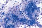
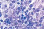
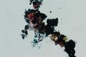
- Divide fluid between an EDTA and 2 plain tubes. Add two drops of formal saline to one plain tube and label. Air dried smears can be made of any thick pieces of mucus (Fig 1)
- Withdraw the endoscope or catheter BAL results in pulmonary oedema, congestion and alveolar collapse and supplemental oxygen should be given throughout the procedure and for 5-20 minutes afterwards. Where possible, oxygen saturation should be monitored.
Please visit www.nwlabs.co.uk or see our current price list for more information


