Castration - Donkey
Introduction
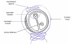
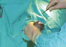
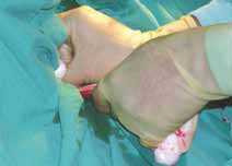
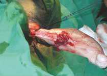
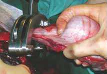
Castration in the donkey is complicated by the small size of the patient necessitating general anaesthesia, and the need for ligation of the spermatic cord. In addition, a wide range of ages may be presented for castration. At The Donkey Sanctuary the range can be from 6 months to 29 years. There are also a number of donkey owners who request castration of foals only a few days old.
In theory, castration of the very young donkey can easily be done under sedation and local anaesthetic, with an assistant holding the foal. In practice, one must bear in mind the stress of the procedure, and perform a full work-up including measurement of IgG before attempting castration. Unless the conditions are very clean there is the risk of the foal succumbing to neonatal sepsis after surgery.
Castration of the mature adult donkey can be complicated by the presence of excess fat within the scrotum and inguinal region. Surgery at this late age is unlikely to remove behavioural problems associated with stallion-like tendencies.
Ideally castration should be performed somewhere between 10 to 18 months old and at a time of year avoiding major fly worry. Tetanus anti-toxin should be administered if there is any doubt about the tetanus vaccination history.
Cryptorchidism occurs in donkeys as in horses. A good examination of the inguinal region should be performed prior to surgery and if in doubt a rig test should be carried out. This involves injection of 6000 iu human chorionic gonadotrophin and sampling before and between 30 to 120 minutes after the injection.
There are three methods of castration used:
- closed
- semi-closed using a scrotal approach
- semi-closed using a cranial inguinal approach
Each method has advantages and disadvantages, and the decision on which method to use is based on surgical experience and size/age of patient.
Castration by the closed technique
This is the method most commonly used in immature and young, slim, adult donkeys. It may not be the best method for mature individuals with significant fat deposits, or sexually active donkeys.
The term ‘closed castration’ refers to the fact that the parietal tunica vaginalis is not opened, and there is, therefore, no direct access to the abdominal cavity.
The surgery is performed under general anaesthesia in the field or in theatre. It is essential that the surgeon is adequately assisted with administering the anaesthetic. The surgery will be longer than for a simple open castration and can be fiddly for those not used to performing it. In addition, donkeys tend to require top-up doses of anaesthetic agents and may wake too quickly if a simple alpha-agonist/ketamine combination is used.
For in-field castration, placement of an intravenous catheter is recommended, and having available top-up doses of a suitable anaesthetic e.g. alpha-agonist/ketamine, triple drip or thiopentone bolus. Antiinflammatory medication is included in the initial induction protocol. Antibiotics, if used, may best be administered pre-operatively.
Once the donkey is recumbent, he is positioned in lateral recumbency with the uppermost hind leg tied to expose the surgical area. After cleaning the site, local anaesthetic (3 to 10 ml) is injected into each testicle. This will reduce the depth of anaesthesia needed and ensure better exposure of the cord, as the donkey is less likely to retract the testicle if it is anaesthetised.
The lower testicle is operated on first, to ensure good visualisation. The skin is incised, but the incision is not carried through the tunica vaginalis communis. A dry swab and blunt finger dissection is the best way to strip the tissue away and expose the spermatic cord and cremaster muscle. It is best to remove fat deposits at this stage as, if left in place, they may protrude through the scrotal wound and wick infection into the wound.
If the donkey is retracting the testicle, an assistant may be useful to hold it and allow good ligature placement. It is important that the assistant does not pull excessively and apply undue tension when the ligatures are tightened. For ligation, five metric absorbable sutures are placed around the cord and tightened slowly and firmly to prevent retraction of the vessels within. Sutures may be anchored in the cremaster muscle or placed as a transfixing ligature, especially in more mature individuals. It is wise to apply tissue forceps to the proximal cord before emasculating; this allows one to check for haemorrhage after emasculation. A standard emasculator is applied 2 cm distal to the ligature and left in place for two minutes.
This procedure is repeated on the upper testicle. The incisions are left open to drain and heal by second intention.
Post-operatively, the donkey must be exercised to prevent swelling and, if necessary, anti-inflammatory drugs should be continued for three to five days.
Castration by the semi-closed technique
In mature individuals with well-developed testes or large fat deposits in the scrotum, there is more risk that ligatures may not adequately crush the structures within the cord if applied in a closed technique. For this reason, one of the semi-closed methods may be chosen. This involves incising through the parietal vaginal tunic and ligation of the contents. There is a greater possibility of introducing infection by this method and, if it is chosen, a high degree of asepsis is required, preferably in a hospital environment.
Semi-closed castration via the scrotal approach
This technique is suitable for quiet, mature jacks where a simple closed technique may not provide adequate haemostasis. A strict standard of asepsis is required.
The donkey is prepared as for the closed castration technique. The tunica vaginalis communis is opened by incision and the neurovascular cord is ligated with 3-metric absorbable sutures, checked for haemorrhage and allowed to retract into the vaginal process. The remaining vaginal process is similarly ligated and the distal portion removed by emasculators or surgical division. It is important to perform this procedure as proximal as possible and to remove all spare fat and fascia that may act as a focus for infection. In mature donkeys there is a good blood supply to the scrotum, and care must be taken with the many vessels in the area, using diathermy or ligation if necessary. While in theory the skin could be closed, the amount of swelling is usually unacceptable without ventral drainage.
Semi-closed castration via the inguinal approach
As it is time-consuming, this technique is only suitable if hospital facilities are available. For a full description see Du Preeze (1999).
It is the most suitable method for large, sexually active donkeys, as there is less risk of infection. We have had experience with large mature jacks post-castration continuing to be sexually active and forcing further lengths of vaginal process down the inguinal canal leading to post-operative infection. In the semi-closed technique via the inguinal approach this is less likely.
The operation is performed with the donkey in dorsal recumbency. An incision is made over the external inguinal ring and the spermatic cord is located and withdrawn. Using gentle but firm traction, the testicle is withdrawn through the incision, thus everting the scrotum. The scrotal ligament is divided, freeing the testicle. The cremaster muscle is separated from the vaginal tunic and ligated. A small incision is then made in the vaginal tunic and the spermatic vessels and ductus deferens are ligated and divided. After checking for haemorrhage, the stump is replaced in the vaginal tunic. Two 5-metric ligatures are then placed around the whole cord and tightened. The testicle and remaining vaginal tunic are then removed. The wound is closed routinely using subcutaneous and absorbable skin sutures.
The donkey needs two days of box rest followed by five days of hand walking twice daily. In our experience this method can result in unacceptable swelling as donkeys are not always easy to hand walk and tend to be very immobile if box rested. If this method is used a good exercise regime must be implemented.
Complications of castration
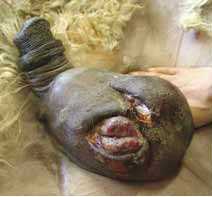
It is good practice to be aware of complications that can occur following castration, and discuss with the owner of the donkey what to look out for in the period following the operation. It is also sensible, if one is operating in the field, to have sufficient equipment and anaesthetics available for correction of haemorrhage should it occur, for example, a spare sterile kit, long artery forceps, sterile packing material, and fluids.
Haemorrhage is the most immediate complication and can be lifethreatening. If pressure from sterile packing is not controlling the flow, the donkey may need to be rapidly re-anaesthetised to locate the source of the haemorrhage. The blood may be from the large scrotal vessels, external pudendal vessels or from the testicular artery. The scrotal wound must be cleaned and the cut ends of the testicular artery located and re-ligated if necessary. The risk of infection occurring after such an emergency is high, and antibiotics will be needed. The donkey must be assessed for blood loss and haemorrhagic shock, and treated accordingly.
Evisceration is unlikely following a closed castration. However, small pieces of fat and fascia may prolapse out of the wound. Every effort should be made to trim these away during surgery; if they are found hanging from the wound, they can act as a route of infection into the wound. Small pieces of tissue may be cut away under sedation if they are fresh; larger pieces may require removal under anaesthetic to ensure asepsis.
Excessive swelling may be a problem in donkeys due to their sedentary nature, especially in older animals. Every effort must be made to encourage exercise and it is advisable to use non-steroidal anti-inflammatory drugs. Hosing the wound can cause waterlogging of the tissues and is not recommended. Uncontrolled swelling can lead to secondary paraphimosis, which may be slow to resolve.
Infection can be superficial and easily dealt with, or deeper, leading to involvement of the vaginal tunic and scirrhous cord. Any suspicion of infection should be promptly investigated under sedation using a gloved hand. Local superficial infection is best dealt with by enlarging the incision sites to improve drainage, combined with cleaning with povidine–iodine and a course of antibiotic treatment. If infection is within the vaginal tunic, repeat surgery is required to resect all affected tissue and this may need to be combined with scrotal ablation if the scrotal tissue is also oedematous and infected.
Nursing Care
Donkey stallions need firm, confident handling. They can be very strong and may require a bit in the mouth when being led. Be aware that they may bite!
The stable door should be high enough so that they cannot see over or attempt to jump it. Some stallions can become very excited by the sight of other equines.
Castration is generally performed under general anaesthetic.
Post-operatively the bed should be kept as clean as possible to help avoid infection. The donkey should receive daily exercise to help prevent and reduce swelling. If he leads well and is not too strong he can be exercised in hand.
If he is turned out, ensure the field is well fenced and there are no other equines in sight. Vigorous exercise the day after surgery should be avoided as it may promote haemorrhage.
| Castration - Donkey Learning Resources | |
|---|---|
 Search for recent publications via CAB Abstract (CABI log in required) |
Donkey castration publications |
 Full text articles available from CAB Abstract (CABI log in required) |
Complications of castration. Adams, S. B.; The North American Veterinary Conference, Gainesville, USA, Large animal. Proceedings of the North American Veterinary Conference, Volume 20, Orlando, Florida, USA, 7-11 January, 2006, 2006, pp 72-73, 3 ref. |
References
- Dabinett, S. (2008) Nursing care In Svendsen, E.D., Duncan, J. and Hadrill, D. (2008) The Professional Handbook of the Donkey, 4th edition, Whittet Books, Chapter 18
- Thiemann, A. (2008) Surgery In Svendsen, E.D., Duncan, J. and Hadrill, D. (2008) The Professional Handbook of the Donkey, 4th edition, Whittet Books, Chapter 16
- Du Preeze, P.M. (1999). ‘Castration - update on techniques’. Proceedings of the Annual Conference of the British Equine Veterinary Association,Newmarket, p 137.
|
|
This section was sponsored and content provided by THE DONKEY SANCTUARY |
|---|
Webinars
Failed to load RSS feed from https://www.thewebinarvet.com/surgery/webinars/feed: Error parsing XML for RSS