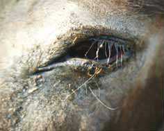
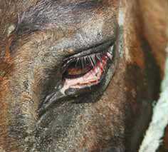
Laceration - Donkey
Definition: a laceration is a traumatic tearing of the skin in an uncontrolled fashion. |
|---|


Lacerations are the commonest skin injuries in donkeys. Multiple tears in the skin may be accompanied by bruising but haemorrhage is rarely a problem.
Management of lacerations usually involves careful wound preparation and restoration of anatomical integrity by means of sutures or staples. Antibiotic therapy is usually applied for a period of four to six days. The absence of ‘spare’ (loose) skin on limbs means that large deficits in these sites require particular care. There is seldom any spare skin that can be mobilized and so a prolonged recovery and/or expensive surgical procedures may be expected.
Degloving injuries are commonest on the upper limb regions. The skin on the lower limb is probably more firmly attached and seldom ‘degloves’ in the same way as the upper limb and body trunk. These injuries should be promptly treated to restore as much of the skin as possible to its original position (even if it is probably non-viable). Degloving of limbs usually involves at least some horizontal skin laceration and is usually in a downward direction so that the skin hangs around the limb. The exposed subcutaneous tissues rapidly become dry and infected but remarkably little bleeding occurs in most such cases. The blood supply to the upper margin of the wound is usually intact and so this is less of a problem than the distal wound margin, which is invariably compromised, especially at the most central part of the wound margin. Sloughing of the skin along this margin is common.
The wound should be irrigated with copious warm sterile saline and protected from further contamination by application of a hydrogel to the exposed tissues. This will minimize dehydration and infection. Restoration of the flap to its normal position as far as possible maintains warmth, prevents further contamination and devitalisation and covers the exposed tissues with a biological dressing. If feasible, a bandage should be applied to limit further damage until a detailed examination can be performed. Movement of the limb should be minimized so that tension on the wound is reduced as far as possible. Shear forces will be maximal during movement of the underlying muscles relative to the skin.
The wound should be carefully examined (possibly even under general anaesthesia) and, after superficial irrigation, all obvious foreign matter and devitalised subcutaneous tissues are scrupulously removed. Deeper injuries are treated by lavage and, if indicated, by suturing the defects with an absorbable suture material of suitable diameter and pattern. Skin should not be removed unless it is totally devitalised and shredded; this applies particularly to the eyelids.
Once the deep tissues have been repaired (possibly with drain placement), the skin is restored to its normal position. Excessive tension on the wound site will almost always result in wound dehiscence and further complications. Carefully placed and securely knotted sub-cutaneous ‘walking’ sutures of a fine absorbable material may allow the wound to be closed without undue tension and will provide support for the flap of skin. However, if the injury is more than one or two hours old, the skin will have shrunk significantly and it may be difficult to restore it to its natural position. Undermining the adjacent attached skin and making small 1 cm long overlapping relieving incisions (running parallel to the wound margins) may permit the skin to be restored fully to the normal site without any undue tension on the wound margin. Alternatively, it is sometimes wise not to attempt to restore the skin fully to its position and simply suture the skin to the underlying tissues, as near as possible to the wound margin without excessive tension. The uncovered area will then heal by second intention but will of course be much smaller than the original wound and may be amenable to skin grafting in due course.
The skin wound is closed using interrupted horizontal or vertical mattress sutures with monofilament nylon (4 or 5 metric/1 or 2 USP). Tension across the wound site can also be relieved by using supported quill sutures or a stent.
Suitable dressings and bandages and antibiotic therapy are useful in all cases. Movement should be restricted depending on the extent and type of wound, and dressings should be changed as and when indicated.
Variable necrosis of at least part of the skin margin is commonly present. In any case the necrotic tissue will eventually need to be removed and the wound allowed to heal by second intention or by some form of grafting.
Uneventful healing should take place regardless of the anatomical location of the wound but can be problematic, especially on the limbs. Complications may be present such as skin loss or degloving and such wounds are best regarded as ‘complicated’. The prognosis is less favourable than for incised wounds because tissue necrosis and sloughing are frequent complications.
References
- Knottenbelt, D. (2008) The principles and practice of wound mamagement In Svendsen, E.D., Duncan, J. and Hadrill, D. (2008) The Professional Handbook of the Donkey, 4th edition, Whittet Books, Chapter 9
|
|
This section was sponsored and content provided by THE DONKEY SANCTUARY |
|---|