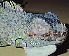
Lizard Dermatitis
Introduction
Skin infections are not uncommon and present clinically with a variety of forms. These include necrotic dermatitis (skin or scale rot) and blister disease (vesicular lesions). This condition is usually due to damp and dirty environmental conditions allowing bacterial and fungal growth; it has also been associated with parasitism (internal and external) and 'stress' (Branch et al., 1998).
Clinical signs
- Blisters with clear or bloody fluid
- Crusts
- Ulceration
- Erythema
- Dark discoloration of skin
Diagnosis
- History
- Clinical examination
- Microscopy, culture and sensitivity: aseptic sampling of unburst blisters and swabbing of sores
- Cytology
- Haematology and biochemistry
Treatment
It should be topical and systemic, and based on sensitivity testing. For extensive sores, dilute chlorhexidine can be used to bathe the animal and soak a paper substrate.
- Debridement
- Antibiotics and creams (silver sulfadiazine)
- Supportive care
| This article is still under construction. |
