Small Animal Orthopaedics Q&A 16
| This question was provided by Manson Publishing as part of the OVAL Project. See more Small Animal Orthopaedics Q&A. |
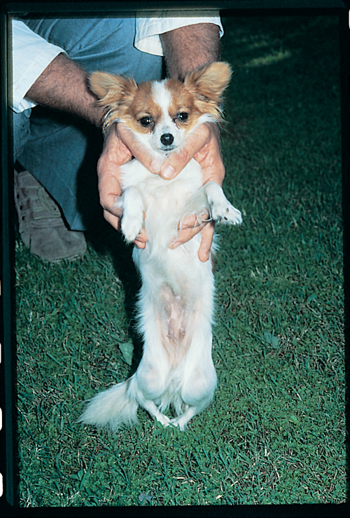
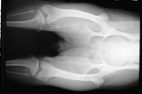
A photograph and a ventro dorsal view radiograph of the pelvis and hindlimbs of a ten-month-old, female Papillon that walks with an extremely crouched hindlimb posture. When purchased at eight weeks of age the dog was able to walk but appeared ‘bow-legged’. As the dog has grown, the dog’s hindlimb gait has become more crouched and the dog’s mobility has decreased. Both stifles are held permanently in flexion and cannot be fully extended. Both hindpaws are internally rotated. Overt pain cannot be elicited on manipulation of the hindlimbs.
| Question | Answer | Article | |
| Describe the radiographic abnormalities. | The abnormalities are similar in both hindlimbs. There is a bowing deformity of the femur with the distal femur bowed medially (genu varum) and hypoplasia of the medial femoral condyle. There is a medial torsional deformity of the proximal tibia of almost 90° with medial deviation of the tibial tubercle. Also present is a tilting of the transcondylar axis of the femorotibial articulation. Both patellae are positioned medial to their respective femoral condyle. The severity of the radiographic abnormalities are consistent with bilateral grade IV medial patellar luxation. |
Link to Article | |
| What other specific musculoskeletal abnormalities would this dog be expected to have on physical examination? | The quadriceps muscle group is responsible for stifle extension and its main antagonist muscle group is the hamstring muscle group (semimembranosus, semitendinosus, biceps femoris muscles). Inability to extend the stifle may arise due to changes within the joint itself, failure of the quadriceps muscle group to extend the joint or restriction of extension caused by failure of the hamstring muscle group to relax. Palpation should include evaluation of these muscle groups and the stifle joint to ascertain the reason the stifle cannot be extended. The location of the patella and tibial tubercle should be determined, as the position of these structures in relation to the axis of the femur and tibia influence the ability of the quadriceps to extend the stifle. In this dog there is mild effusion and joint capsule thickening in both stifles and mild, generalized hindlimb muscle atrophy. The distal portion of the quadriceps muscle group deviates to the medial aspect of the limb and the patella is permanently fixed medial to the trochlear groove. The tibial tubercle is positioned on the medial side of the stifle and the distal limb rotation appears to originate at a point located just distal to the stifle. |
Link to Article | |

