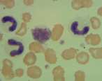
Leucocyte pattern recognition
Leucocyte numbers can change rapidly. Patterns of change can help to determine the underlying cause.
Adrenaline mediated leucocytosis
This is a transient, physiological change caused by the release of adrenaline,leading to the rapid shift of cells from the marginating pool into the circulating pool. This is most often seen in young excitable cats. There is leucocytosis with neutrophilia, often with lymphocytosis.
Corticosteroid mediated leucocytosis
This pattern of change may be associated with endogenous corticosteroids due to stress or hyperadrenocorticism, or exogenous steroids. Classically there are five changes seen in dogs: leucocytosis with neutrophilia, monocytosis, lymphopaenia and eosinopaenia. Not all changes are always present, neutrophilia and monocytosis are the most common. The leucocytosis is usually mild (20-25 x109/l) but particularly after steroid therapy can increase to 30-40 x109/l. In cats monocytosis is rarely a feature of a steroid response.
Inflammation
Leucocytosis and particularly neutrophilia may be a sign of inflammation. Leucocyte changes vary according to the severity and duration of inflammation and ability of the animal to respond to the inflammatory stimulus.
Neutrophilia
Initially an acute inflammatory stimulus, mediated through cytokines, causes release of the storage pool of mature neutrophils from the bone marrow. If the stimulus persists band neutrophils, metamyelocytes and occasionally myelocytes and promyelocytes are released from the bone marrow granulocyte pool. This is evident in the peripheral circulation a left shift.
A left shift of >1.0 x10⁹/l band cells is diagnostic for inflammatory disease. Milder left shifts (0.3-1.0 x10⁹/l bands) may occur with haemorrhage, chronic or granulomatous inflammation.
Neutrophils less mature than band cells indicate increasing intensity of an inflammatory response. The greatest left shift is seen in early disease when there may also be a stress lymphopaenia. Myeloid hyperplasia in the bone marrow becomes established over several days until production and tissue demand are similar. As this happens the neutrophilia becomes more mature, the left shift diminishes or may disappear. As inflammation becomes chronic, lymphocytosis and monocytosis may develop, however, there may be no overall leucocytosis. Mature neutrophilia (>40 x109/l in dogs and >30 x109/l in cats) indicates a significant inflammatory process.
Distinguishing between inflammation and corticosteroid response
This can be problematic and is not always possible. A left shift supports inflammation. Serum protein concentrations and serum protein electrophoresis can be useful. During serum protein electrophoresis plasma proteins are separated into albumin and globulin fractions (α1, α2, β1, β2, and γ globulins). α and β globulins are increased with acute inflammation. Polyclonal increases in the gamma fraction support established inflammation.
Prognostic indicators in inflammation
Degenerative left shift. Tissue demand exceeds the rate of neutrophil production within the bone marrow, leaving insufficient time for cell maturation. There are two definitions; the first is the most common.
- Band cells (nonsegmented neutrophils) exceed mature neutrophils regardless of the white cell count
- A significant left shift (>1.0 x10⁹/l band cells), or band cells represent >10% of leucocytes, with leucopaenia/neutropaenia
Leucopaenia. Tissue demands exceed the rate of bone marrow production of leucocytes.
Leukemoid reaction. There is a marked leucocytosis with neutrophilia (>50-100 x109/l). This occurs with localized inflammation or infection for example pyometra, abscess or peritonitis. A leukemoid reaction may be seen with IMHA; the destruction and phagocytosis of erythrocytes appears to be a strong stimulus for inflammation.
Toxic neutrophils. Indicate accelerated granulopoiesis in response to an inflammatory stimulus. Often associated with gram negative sepsis.
Severe or persistent lymphopaenia. Indicative of severe or persistent stress or immunodeficiency.
Leucoerythroblastic reaction
This occurs when diseases such as myelofibrosis, marrow necrosis or neoplasia disrupt the bone marrow-blood barrier, allowing the premature release of haematopoietic cells into the blood. Immature erythroid and myeloid cells appear in the circulation mimicking erythroleukaemia.
Neutropaenia
Excessive tissue demand for neutrophils. In acute, intense inflammation; usually followed by a rebound neutrophilia with a left shift; a repeat haemogram in 72 hours may clarify.
Decreased neutrophil production. Viral infections such as feline panleucopaenia virus infection and FeLV. Oestrogens toxicity (exogenous or endogenous from an ovarian cyst or a Sertoli cell tumour), cytotoxic drugs, phenylbutazone, phenobarbitone, chloramphenicol and griseofulvin.
Ineffective production. Myeloid leukaemia or myelodysplastic syndrome (MDS). In cats the latter is usually associated with FeLV infection.
Monocytosis
Often seen, with neutrophilia, as a stress or corticosteroids response in the dog. In cats monocytosis is rarely seen following exposure to corticosteroids and is most often seen with inflammation.
Monocytosis can also occur in any disease where there is increased demand for macrophages (removal of necrotic debris, phagocytosis of abnormal red cells, granulomatous diseases etc).
Although macrophages increase in tissue fairly late in an inflammatory process, monocytosis can occur with both acute and chronic disease. Extreme monocytosis may indicate leukaemia but is rare.
Eosinophilia
Most eosinophils are in the tissue pool, particularly associated with the skin, respiratory and alimentary tracts. The blood to tissue ratio is approximately 1:300. Eosinophilia indicates a site of eosinophilic inflammation although even intense eosinophilic inflammation is not necessarily associated with eosinophilia. Eosinophilia may be seen with parasitic disease and hypersensitivity responses. A rebound mild eosinophilia may be seen following withdrawal of steroid therapy and with hypoadrenocorticism. Neoplasia such as mast cell tumours and lymphoma in cats can be associated with eosinophilia. Eosinophilic leukaemia is rare, poorly documented and difficult to differentiate from hypereosinophilic syndrome in cats when mature eosinophils infiltrate internal organs.
Eosinopaenia
Response to stress or corticosteroids
Basophilia
Basophils are uncommon in the peripheral blood of dogs and cats. Their presence may indicate a hypersensitivity response or parasitism with tissue invasion.
Lymphocytosis
Mild lymphocytosis may be seen in cats as an adrenaline response. Persistent lymphocytosis may reflect antigenic stimulation due to viraemia, chronic infection, immune mediated disease including hypersensitivity disorders, primary IMHA in cats and recent vaccination. In these cases reactive lymphocytes may be seen. Transient or reactive lymphocytosis must be differentiated from lymphocytosis due to lymphoid neoplasia (chronic lymphocytic leukaemia, acute lymphoblastic leukaemia or leukaemic lymphoma). Lymphocyte morphology and the magnitude of the lymphocytosis may help to determine the cause.
Lymphopaenia
Frequently seen as a stress (physiological, pathological) or corticosteroid response (exogenous or HAC). Other considerations include viral disease, immunodeficiency and loss of lymphocytes from circulation (chylothorax - particularly if repeatedly drained and lymphangiectasia).
Authors & References
NationWide Laboratories#In Partnership with NationWide Laboratories
