File:478px-Bacteria cell wall svg- franciscosp2.png
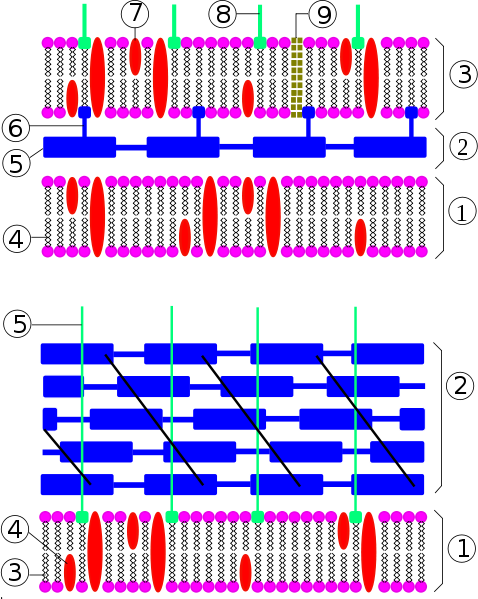
Revision as of 12:21, 3 September 2008 by Eayton (talk | contribs) (Bacterial cell wall. Top: Gram-negative cell wall. 1-inner membrane, 2-periplasmic space, 3-outer membrane, 4-phospolipid, 5-peptidoglycan, 6-lipoprotein, 7-protein, 8-LPS, 9-porins. Botton: Gram-positive cell wall. 1-cytoplasmic membrane, 2-peptidoglycan)
478px-Bacteria_cell_wall_svg-_franciscosp2.png (478 × 599 pixels, file size: 121 KB, MIME type: image/png)
Bacterial cell wall. Top: Gram-negative cell wall. 1-inner membrane, 2-periplasmic space, 3-outer membrane, 4-phospolipid, 5-peptidoglycan, 6-lipoprotein, 7-protein, 8-LPS, 9-porins. Botton: Gram-positive cell wall. 1-cytoplasmic membrane, 2-peptidoglycan, 3-phospholipid, 4-protein, 5-lipoteichoic acid. Copyright Franciscosp2 2008
File history
Click on a date/time to view the file as it appeared at that time.
| Date/Time | Thumbnail | Dimensions | User | Comment | |
|---|---|---|---|---|---|
| current | 10:08, 20 July 2010 |  | 478 × 599 (121 KB) | Eca02csb (talk | contribs) | {{Information |Description=Bacterial cell wall. Top: Gram-negative cell wall. Bottom: Gram-positive cell wall. |Source=WikiMedia Commons |Date=03/05/08 |Author=Franciscosp2 |Permission=See above }} |
You cannot overwrite this file.
File usage
The following page uses this file:
