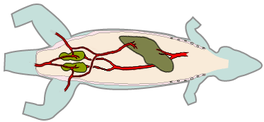
Lizard Cardiovascular System
| This article is still under construction. |
Heart
Lizards have a three chambered heart with left and right atria and a single ventricle - the interventricular septum is only partially complete but serves to channel deoxygenated blood to the pulmonary trunk and oxygenated blood to the circulation. In addition the atria contract at different times, thus directing the flow of blood between the sponge-like arranged muscle bundles which form the major part of the ventricle.There are paired left and right aortic arches which join caudal to the heart to form a single dorsal aorta.
The renal portal system
Reptiles have a renal portal system where venous return from the tail and hindlimbs may be filtered through the kidneys to bathe the renal tubules. Venous return from the tail and hindlimbs may be filtered through the kidney or liver. When filtered through the kidney, the blood bathes the renal tubules.
- The injection of drugs that are cleared via tubular excretion into the caudal half of the body could result in lower than anticipated serum concentration because of their excretion in the urine before entering the systemic circulation. This could potentially result in increased renal toxicity when using nephrotoxic drugs (eg. aminoglycosides); however the results of limited pharmacokinetic studies suggest that the renal portal system has less effect on drug uptake and distribution than previously thought. Furthermore, shunts exist that carry blood directly from the renal portal system to the postcava, bypassing the renal parenchyma.
Surgery
- Of surgical importance is the large ventral abdominal vein that lies along the inner surface of the abdominal wall in the midline. It must be avoided in a celiotomy incision, for example by using a paramedian incision. It should also be protected with saline solution-moistened gauze and it can be ligated without clinical effect.
- Venipuncture can be done with the ventral caudal vein in species that do not undergo tail autotomy, with the axillary venous plexus or with the jugular vein.
- Intravenous catheters can be placed in the cephalic or jugular vein of large specimens (with long necks), with a cutdown technique, or in the ventral caudal vein using a lateral approach aiming craniad. This route can be used to give IV fluids, as well as through an intraosseous catheter placed in the femur (distal end) or tibia.
For more information on reptile surgery, see Lizard and Snake Surgery.
References
- Mader, D.R. (2005). Reptile Medicine and Surgery. Saunders. pp. 1264. ISBN 072169327X
