| This article is still under construction. |
Introduction
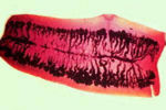
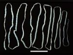
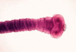
Like most adult tapeworms, Taenia species live in the small intestine. The different species vary in length from 0.5-15m long. Identification is based on the hooks on the scolex. Despite their impressive size, the adult tapeworms of little clinical significance. The metacestodes of some species, however, may cause disease or meat inspection losses. The human pork tapeworm, Taenia solium, is a dangerous zoonosis but, fortunately, it does not occur in the UK.
| Taenia | Final Host | Intermediate Host | Metacestode | Obsolete Name for Metacestode |
|---|---|---|---|---|
| T. saginata | Human | Cattle | Cysticercus in muscle | C. bovis |
| T. solium | Human | Pig | Cysticercus in muscle | C.cellulosae |
| T. ovis | Dog | Sheep | Cysticercus in muscle | C. ovis |
| T. hydatigena | Dog | Sheep etc. | Cysticercus in peritoneum | C. tenuicollis |
| T. pisiformis | Dog | Rabbit | Cysticercus in peritoneum | C. pisiformis |
| T. multiceps | Dog | Sheep | Coenurus (various sites) | C. cerebralis |
| T. serialis | Dog | Rabbit | Coenurus (various sites) | C. serialis |
| T. taeniaeformis | Cat | Mouse etc. | Strobilocercus in liver | S. fasciolaris |
Nomenclature of Metacestodes
The preferred nomenclature is, for example, “the metacestode of Taenia hydatigena”.
Taeniid Eggs
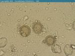
Have a striated “shell”, and are small (half the size of a strongyle egg). The six hooks of the tapeworm larva (oncosphere) are sometimes visible inside the egg. One or more gravid segments, each containing some 150,000-250,000 eggs, are shed daily and pass out of the anus. Eggs are released into the environment by the motile segments.
Taenia spp of Humans
T. saginata, the Beef Tapeworm of Humans
The adult in the human small intestine is unarmed, that is, no hooks on the scolex. Humans are infected by eating undercooked beef. Cattle are infected by ingesting eggs on pasture, or from contaminated utensils (e.g. calves from the milk-bucket). Transmission:
a) Direct: eggs reach pasture by direct deposition of human faeces
b) Indirect: by use of sewage sludge on agricultural land
c) Birds: may transport whole proglottids from effluent outlets or, if they swallow the segment, pass the eggs with their droppings.
Cysticerci
Grow to approximately 1cm long in the bovine intermediate host. If acquired during calfhood = survive; if acquired in later life eventually die = caseous = calcified. May be found in any striated muscle, but the highest densities are in heart and masseters.
Epidemiology
May differ from place to place. For example: - In the UK, very low prevalence in human population + generally high standards of sanitation = low transmission rate to cattle = low general prevalence in cattle = no herd immunity = sporadic “cysticercoid storms”. - In East Africa, high prevalence in human population + lack of sanitation in rural areas = high rate of transmission to cattle = many calves infected = calfhood infection persists, but animals immune to reinfection.
T. solium, the Pork Tapeworm of Humans
The adult tapeworm, which occurs in the human small intestine, has hooks on the scolex. The life-cycle is similar to that of T. saginata, except that the pig is the intermediate host. Heavy infections in pigs may be detected in vivo by inspecting the tongue for cysticerci. T. solium is particularly prevalent in communities where pigs are allowed to scavenge freely around human habitation. This species is particularly dangerous as the cysticerci (known as ‘measles’), which normally occur in the pig, can also develop in human tissues (brain and musculature). This may happen by swallowing eggs from the environment or, in people carrying a tapeworm, as a result of eggs being brought forward in the alimentary tract by retro-peristalsis.
Taenia spp of the Dog
- Several species occur in dogs, varying in length from approximately 0.5-5m.
- Some species may also occur in the fox.
- The adults live in the small intestine and appear to cause little harm, but the exiting proglottids may cause pruritis.
- The prevalence of each species varies in different groups of dogs depending on their diet, that is, whether or not they have access to fresh meat, offal or rabbits.
- The prepatent period is generally 6-8weeks.
- It is important to differentiate the gravid proglottids of Taenia from those of Dipylidium. Taenia segments:
- are rectangular
- there is only one lateral genital pore
- the eggs are single (i.e. not in packets like Dipylidium)
T. ovis
- Cysticerci occur in the musculature of sheep = condemnation of meat on aesthetic grounds.
- Common in some sheep-raising areas of the UK.
- Known as ‘sheep measles’ in Australasia, where it causes economic loss to the export industry.
- Consequently, a recombinant oncosphere antigen vaccine is available in New Zealand.
T. hydatigena
- The commonest species in the UK.
- Sheep are the most frequent intermediate host, but the cysticerci can establish in other animals.
- The oncosphere hatches out of the egg in the small intestine of the sheep.
- Oncospheres travel to the hepatic portal system, where they transform to cysticerci.
- The grow rapidly while migrating through liver parenchyma, then to the peritoneal cavity.
- The cysticerci in the peritoneal cavity are approximately 8cm long; often found adhering to the omentum.
- Usually, liver damage heals, forming fibrotic tracts, which leads to condemnation at meat inspection.
- If a sheep swallows a whole proglottid, it leads to liver damage, and ultimately death (“cysticercosis hepatica”), but this is a rare event affecting a single animal in a flock.
T. pisiformis
- It is similar to T. hydatigena, except that the cysticerci are pea-sized and are found on the omentum of rabbits.
T. multiceps (also known as Multiceps multiceps)
- Widespread distribution in the UK; particularly common in parts of mid-Wales.
- The eggs hatch in the small intestine of the sheep.
- The oncosphere enters the blood stream and travels to the brain, where it migrates through brain tissue.
- The metacestode (a coenurus) occurs inside the skull, lying on the surface of the brain.
- It forms a 5cm long space occupying lesion.
- This leads to neurological signs, including gid – circling, ataxia and blindness.
- It also causes softening of the skull above the lesion.
- If a whole proglottid is ingested, it can lead to acute encephalitis (very rare).
T. serialis
- The coenurus forms in intermuscular connective tissues of rabbits, often causing a soft subdermal swelling.
- Cases in pet rabbits probably originate from eggs shed by urban foxes.
Taenia spp of the Cat
T. taeniaeformis
- The metacestode appears as a pea-sized nodule in the liver of mice and other small rodents.
- Cats will continue to become re-infected if they are hunters.
Peritoneal Cavity Parasitic - Pathology inhabited by Taenia hydatigena, Taenia pisiformis, Taenia ovis
- Taenia solium and T.ovis, T. saginata, Multiceps serialis in myositis


