Category:Image Review Completed
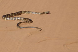
Revision as of 14:09, 21 October 2010 by Suzannah.stacey (talk | contribs) (Created page with "250px|thumb|right|'''Snake locomotion: sidewinding''' Source: Wikimedia Commons; Author: José Reynaldo da Fonseca (2007)")
Media in category "Image Review Completed"
The following 65 files are in this category, out of 65 total.
- 763px-Cryptosporidium parvum 01.jpg 763 × 600; 39 KB
- Adrenal Gland Schematic..jpg 903 × 508; 39 KB
- Ameloblast Histology.jpg 800 × 560; 70 KB
- AscendingReticularFormation.jpg 589 × 333; 11 KB
- Astrocyte.jpg 800 × 532; 75 KB
- Body Compartments.jpg 631 × 253; 17 KB
- Calfvssheepblood.jpg 960 × 720; 73 KB
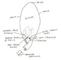
- Canine Auricular Cartilages.jpg 1,280 × 1,264; 228 KB
- Canine Brain CS showing Hypothalamus1.jpg 761 × 496; 50 KB
- Canine Ear Canal.jpg 826 × 584; 61 KB
- Caninelateralventricles.jpg 938 × 529; 52 KB
- Carotidretesheep.jpg 1,130 × 551; 52 KB
- Central Vestibular Pathways.jpg 680 × 400; 35 KB
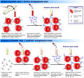
- Coombs test schematic.png 641 × 600; 246 KB
- Developing Eye.jpg 693 × 400; 49 KB
- Donkey5.png 200 × 330; 107 KB
- DSC01703.JPG 3,264 × 2,448; 1.33 MB
- DSC01713.JPG 2,920 × 2,244; 788 KB
- DSC01727.JPG 3,240 × 2,088; 958 KB
- DSC01788.JPG 3,264 × 2,448; 1.05 MB
- DSC01793.JPG 2,592 × 2,092; 932 KB
- DSC01794.JPG 3,168 × 2,092; 846 KB
- Egyptian spiny tail lizard.jpg 800 × 570; 178 KB
- Extrapyramidal system.jpg 562 × 412; 17 KB
- Eye gross structure.JPG 610 × 539; 66 KB
- Gila monster.jpg 800 × 593; 210 KB
- Greentree monitor london.jpg 3,264 × 2,448; 883 KB
- Histology of the Adrenal Glands Medulla..jpg 676 × 387; 47 KB
- Histology of the Adrenal Glands showing zones..jpg 674 × 370; 53 KB
- Histology of the Adrenal Glands..jpg 676 × 379; 48 KB
- Iris in detail.jpg 700 × 408; 62 KB
- Iris.jpg 691 × 400; 36 KB
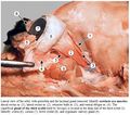
- LH Bursa Cloaca Photo.jpg 601 × 272; 52 KB
- LH Lymph Node Follicles Histology.jpg 666 × 401; 132 KB
- LH Lymph Node Gross Histology.jpg 745 × 546; 122 KB
- LH Lymph Node HEV Histology.jpg 898 × 452; 135 KB
- Nervestructure.jpg 444 × 823; 159 KB
- Pyramidalsystem.jpg 589 × 333; 9 KB
- Rabbit ears.jpg 800 × 600; 118 KB
- Retina numbered.jpg 692 × 441; 38 KB
- Retina of the Dog.jpg 684 × 447; 65 KB
- Section through Cochlea - Histology.jpg 695 × 400; 35 KB
- Spiny tailed lizard enclosure.jpg 2,692 × 2,316; 840 KB
- Stroma.jpg 725 × 462; 48 KB
- The Ascending Pathways.jpg 589 × 333; 9 KB
- TheSpinocerebellarTract.jpg 589 × 333; 8 KB
- Thomes' Fibres Histology.jpg 807 × 612; 98 KB
- Unilateral Vestibular Signs.jpg 640 × 400; 31 KB
- Uromastyx flavifasciata.JPG 2,988 × 2,244; 839 KB
- Vestibular Receptors and Balance.jpg 676 × 400; 56 KB
- WIKIVETautonomicganglion.jpg 663 × 394; 49 KB
- WIKIVETbraindifferentiation.jpg 785 × 429; 28 KB
- WIKIVETcerebellum.jpg 650 × 378; 35 KB
- WIKIVETcerebrum.jpg 631 × 377; 44 KB
- WIKIVETcrosssectionofspinalcord.jpg 635 × 416; 26 KB
- WIKIVETdorsalrootganglion.jpg 652 × 401; 53 KB
- WIKIVETformationofneuraltissue.jpg 793 × 447; 31 KB
- WIKIVETmyeinatednerveinsmallfibrelipdstained.jpg 676 × 347; 37 KB
- WIKIVETmyelinatednervesinsmallfibreinlongittudinlsection.jpg 683 × 346; 32 KB
- WIKIVETmyleinatednerveinsmallfibre.jpg 677 × 360; 34 KB
- WIKIVETneuraltubeformation.jpg 562 × 273; 16 KB
- WIKIVETperipheralnervestructure.jpg 635 × 416; 40 KB
- WIKIVETspinalcord1.jpg 680 × 431; 56 KB
- WIKIVETspinalcord2.jpg 700 × 410; 49 KB
- WIKIVETspinalcord3.jpg 674 × 360; 32 KB


