File:Cyclospora.jpg
Cyclospora.jpg (377 × 347 pixels, file size: 21 KB, MIME type: image/jpeg)
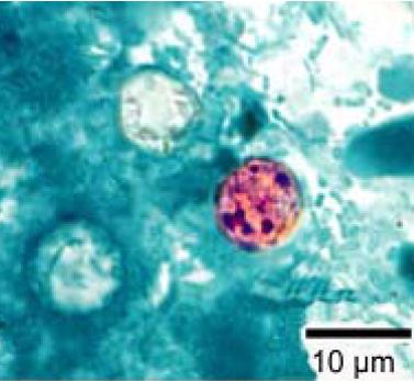
This photomicrograph of a fresh stool sample, which had been prepared using a 10% formalin solution, and stained with modified acid-fast stain, revealed the presence of four en:Cyclospora cayetanensis oocysts in the field of view. Compared to wet mount preparations, the oocysts are less perfectly round and have a wrinkled appearance due to this method of fixation. Most importantly, the staining is variable among the four oocysts. Cyclospora infects the small intestine, and usually causes watery diarrhea with frequent, sometimes explosive bowel movements. Other symptoms can include loss of appetite, substantial loss of weight, bloating, increased gas, stomach cramps, nausea, vomiting, muscle aches, low-grade fever, and fatigue. Some people with Cyclosporiasis do not show any symptoms.
http://en.wikipedia.org/wiki/Image:Cyclospora_cayetanensis_stained.jpg
File history
Click on a date/time to view the file as it appeared at that time.
| Date/Time | Thumbnail | Dimensions | User | Comment | |
|---|---|---|---|---|---|
| current | 18:05, 23 November 2008 | | 377 × 347 (21 KB) | Nabrown (talk | contribs) | This photomicrograph of a fresh stool sample, which had been prepared using a 10% formalin solution, and stained with modified acid-fast stain, revealed the presence of four en:Cyclospora cayetanensis oocysts in the field of view. Compared to wet mount pr |
You cannot overwrite this file.
File usage
The following file is a duplicate of this file (more details):
- File:Cyclospora cayetanensis stained.jpg from Wikimedia Commons
The following 2 pages use this file: