File:Equine protozoal myeloencephalitis.jpg
Equine_protozoal_myeloencephalitis.jpg (329 × 336 pixels, file size: 151 KB, MIME type: image/jpeg)
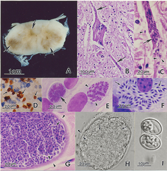
Sarcocystis neurona stages and lesions.
(A). Cross section of spinal cord of horse with focal areas of discoloration (arrows) indicative of necrosis. Unstained.
(B). Section of spinal cord of a horse with severe EPM. Necrosis, and a heavily infected neuron (arrows), all dots (arrows) are merozoites. H and E stain .
(C). Higher magnification of a dendrite with numerous merozoites (arrows). One extracellular merozoite (arrowhead) and a young schizont (double arrowhead).
(D). Section of brain of an experimentally-infected mouse stained with anti-S. neurona antibodies. Note numerous merozoites (arrows).
(E). Immature schizonts in cell culture. A schizont with multilobed nucleus (arrow) and a schizont with differentiating merozoites (arrowheads). Giemsa stain.
(F). Mature sarcocysts with hairlike villar protrusions (double arrowheads) on the sarcocyst wall. H and E stain.
(G). Mature live sarcocyst with numerous septa (arrows) and hairlike protrusions on the sarcocyst wall (double arrowheads). Unstained.
(H). An oocyst with two sporocysts each with banana-shaped sporozoites. Unstained.
http://commons.wikimedia.org/wiki/File:Equine_Protozoal_Myeloencephalitis.jpg
Wikimedia Commons
File history
Click on a date/time to view the file as it appeared at that time.
| Date/Time | Thumbnail | Dimensions | User | Comment | |
|---|---|---|---|---|---|
| current | 14:27, 21 December 2008 | | 329 × 336 (151 KB) | Nabrown (talk | contribs) | Sarcocystis neurona stages and lesions. (A). Cross section of spinal cord of horse with focal areas of discoloration (arrows) indicative of necrosis. Unstained. (B). Section of spinal cord of a horse with severe EPM. Necrosis, and a heavily infected neu |
You cannot overwrite this file.
File usage
The following 2 files are duplicates of this file (more details):
- File:Equine Protozoal Myeloencephalitis.jpg
- File:Equine Protozoal Myeloencephalitis.jpg from Wikimedia Commons
The following 2 pages use this file: