Hoof - Horse Anatomy
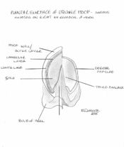
As outlined above, the equine hoof can be divided into three topographical regions; the wall, the frog and the sole. The wall forms the medial, lateral and dorsal aspect of the hoof and it can be further divided into the toe, quarters and heels. At the heel the walls reflect back on themselves at a point called the angles and in doing so forms the bars. The bars, although moving cranially, gradually fade along the edge of the frog and never actually meet. The frog sits between the bars and has an apex facing distally, with 2 crura flanking a central sulcus. Between the crus and bar of each half of the sole lies the collateral sulcus. Opposite the apex, the frog expands forming the bulbs of the heel. The sole is the area distal to the bars and apex of the frog enclosed by the hoof wall. The area where the bars and wall enclose it is known as the angle of the sole.
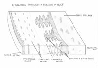
The dermis of the distal phalanx is arranged in hundreds of leaves or laminae, each of which has microscopic secondary laminae. The coronary region has a germinative layer associated with papillae that is responsible for producing the horn tubules that make up the hoof wall. This wall glides distally at a rate of 5-6mm a month and by forming epidermal laminae itself it interdigitates with the underlying dermal laminae. Neither of these laminae are pigmented so when the epidermal laminae appear on the solar surface, a non-pigmented region known as the white line appears. The white line is used as important landmark in farriery as structures central to the line will be dermal and so vascular and sensitive.
The dermis in the frog is also arranged in papillae and produces incompletely keratinised flexuous horn tubules resulting in a soft, elastic horn. The hypodermis of the region of the frog forms the digital cushion. This lies between the ungual cartilages and is collagenous, elastic tissue infiltrated by adipose tissue. At the bulbs of the heel, it is subcutaneous and is soft and loose in texture. The sole area also has papillae that produces superficially flakey horn. The coronary part of the wall is surrounded by a bony prominence called the periople. This soft, lightly coloured area is restricted to this proximal area and is produced by the germative layer covering the papillae. The rest of the hoof is covered by the tectorial layer, this is a very thin layer of horn that is covered distally by the growth of the horn.
A well-trimmed foot should weight bear on its walls, bars and frog. This occurs as the weight applied to the distal phalanx is then transferred across the interdigitating laminae to the hoof wall. Thus an injury resulting in damage to the laminae is of extreme importance to the horse.

