Anatomy of the Enamel Organ
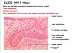
The main components which form the enamel organ are:
- Outer epithelium
- Stellate reticulum- star shaped cells lying between the outer and inner epithelial layers. It has the appearance of connective tissue but is of epithelial derivation.
- Inner epithelium which becomes the enamel secreting ameloblast layer
The enamel organ has many different components. These consist of:
Crown
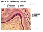
The crown is covered by enamel. It meets the root at the cemento-enamel junction (CEJ).
The crown of incisors have only one cusp. The crown of molars have up to 4 cusps for the grinding of food.
The main cells of the enamel organ are:

