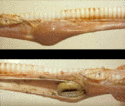
Impaction of the Oesophagus
Also known as: Choke
Introduction
Impaction (obstruction) of the oesophagus or 'choke' is the most common oesophageal disease of cattle and horses. Obstruction usually occurs at the level of the thoracic inlet, the base of the heart or the hiatus oesophagus of the diaphragm (i.e. the narrowest points). In horses, causes of choke include consumption of unsoaked sugarbeet, rapid ingestion of roughage (hay), indadequate mastication of food, or rarely the ingestion of a foreign body (e.g. corn cob). Horses that are fed incorrectly or have poor dentition are more prone to developing the condition.
In cattle, choke usually occurs due to the ingestion of a single solid vegetable or fruit such as a potato, turnip or corn cob.
Clinical Signs
Horse
Although the presentation of a horse with choke may be dramatic, it does not constitute an emergency. The typical sign of choke is dysphagia with regurgitation of food and saliva through the nostrils. Acute onset pain may manifest through alterations in posture, such as an arched neck or abducted elbows. The horse may appear anxious, grunt frequently and make repeated attempts to swallow. If the condition has become long-standing, foetid breath may be apparent. If accompanied by pyrexia, this may indicate the development of aspiration pneumonia.
Cow
Oesophageal obstruction in cattle is a more serious condition than in the horse. Obstruction may result in a failure to eructate leading to the development of free-gas bloat. The clinical signs are similar to those seen in the horse including ptyalism, coughing, arching of the neck, dysphagia and nasal discharge. The expanded rumen causes increased pressure on the diaphragm, reduced venous return to the heart and may lead to respiratory distress or asphyxia. Grinding of the teeth, hypersalivation or protrusion of the tongue may also be seen.
Diagnosis
An initial diagnosis of choke in any animal may be suspected if the above clinical signs are present with a history of acute onset pain and access to unsuitable food. If the obstruction has led to the accumulation of food material in the oesophagus, a mass may be palpable on the left ventrolateral aspect of the neck although this may be difficult to detect in the adult cow. Confirmation of the diagnosis may be achieved by the inability to pass a nasogastric tube or direct visualisation of the obstruction using endoscopy. Other tests that may provide useful information include radiography and ultrasound.
Treatment
Horse: In the horse, treatment is usually conservative as most obstructions resolve spontaneously or with medical treatment. Treatment comprises passage of a nasogastric tube followed by intraluminal oesophageal lavage. Before attempting this procedure it is important to ensure that the horse is well sedated (with its head below the thoracic inlet) so that the risk of aspiration is minimised. The administration of oxytocin may be beneficial, particularly in animals with cranial obstructions. Following removal of the obstruction, analgesia may be provided using a non-steroidal anti-inflammatory drug such as flunixin. Possible recurrence of an obstruction may be avoided by withholding dry food for 72 hours. Initially, fluids only should be offered, followed by gradual introduction of soft foods such as bran mash. Small amounts of hay or haylage may be added gradually. Following treatment of the impaction, it may be beneficial to perform an endoscopic exam of the oesophagus. This aids in identifying any areas of inflammation or ulceration that may require further treatment.
If conservative treatment fails to resolve the problem, surgery to remove the obstruction may be required. Careful consideration should be given before embarking such treatment due to the high complication rate associated with oesophageal surgery.
Cattle: In cattle, rumenal bloat caused by the obstruction is an emergency and requires immediate treatment. This is achieved by trocharisation through the left paralumbar fossa. Once the bloat has been relieved, the obstruction may be manually broken down via percutaneous massage, or may resolve spontaneously due to the large volume of saliva present. As in the horse, a sedative such as xylazine may be administered combined to provide both sedation and muscle relaxation. Broad spectrum antibiotics should be administered if there is any suspicion of aspiration pneumonia.
In cattle, stomach tube can be used to push the obstructive material down to the rumen. For this procedure, keep the animal head at a 45* angle and place the lubricated stomach tube in the oral cavity and slowly move further down the left side of the neck. Care should be taken not to place the tube inside the trachea. (If the tube enters the trachea accidentally, the operator can notice the wind blowing sound, condensation of the tube if it is transparent and the cow will also show dyspnea. The tube should be withdrawn immediately and correct placement into the oesophagus attempted.) When the tube in the oeasophagus touches the foreign body, resistance can be felt. An attendant standing on the left side of the cow can feel the tube end and the foreign body. Once reached at the particular level, gently push the tube further down to move the foreign body. If the foreign body moves down to the rumen, the operator can see the bloat reducing within few minutes and the animal feels more comfortable. (The tube should be lubricated with any available non toxic lubricants before insertion.)
Prognosis
In the horse, the prognosis for a complete recovery after an episode of simple choke is good. In the cow, the prognosis is good providing minimal manipulation and tissue damage has occurred. A poorer prognosis is associated with prolonged obstruction or perforation of the oesophagus.
| Impaction of the Oesophagus Learning Resources | |
|---|---|
To reach the Vetstream content, please select |
Canis, Felis, Lapis or Equis |
 Search for recent publications via CAB Abstract (CABI log in required) |
Oesophageal obstruction publications |
References
- Hillyer, M. (1995) Management of oesophageal obstruction ('choke') in horses In Practice November 1995, 17:450-457
- Hance, S. R., Noble, J., Holcomb, S, Rush-Moore, B. and Beard, W (1997) Treating Choke with Oxytocin Proceedings of the Annual Convention of the AAEP 1997, 43: 338-339
- Haskell, S. R. R. (2008) Blackwell's Five Minute Veterinary Consult: Ruminant Wiley-Blackwell
- Merck & Co (2008) The Merck Veterinary Manual (Eighth Edition) Merial
| This article has been peer reviewed but is awaiting expert review. If you would like to help with this, please see more information about expert reviewing. |
Webinars
Failed to load RSS feed from https://www.thewebinarvet.com/gastroenterology-and-nutrition/webinars/feed: Error parsing XML for RSS
