Difference between revisions of "Peyer's Patches - Anatomy & Physiology"
| Line 1: | Line 1: | ||
| + | {{OpenPagesTop}} | ||
[[Image:LH Ileal Peyer's Patch Diagram.jpg|thumb|250px|right|<p>'''Diagram: Peyer's patch'''</p><sup>©Nottingham Uni 2008</sup>]] | [[Image:LH Ileal Peyer's Patch Diagram.jpg|thumb|250px|right|<p>'''Diagram: Peyer's patch'''</p><sup>©Nottingham Uni 2008</sup>]] | ||
==Introduction== | ==Introduction== | ||
| Line 27: | Line 28: | ||
<br><br> | <br><br> | ||
{{Jim Bee 2007}} | {{Jim Bee 2007}} | ||
| + | |||
| + | {{OpenPages}} | ||
[[Category:Mucosal Associated Lymphoid Tissue]] | [[Category:Mucosal Associated Lymphoid Tissue]] | ||
Revision as of 18:28, 29 June 2012

Introduction
Also called aggregated nodules
Peyer’s patches are lymphoid tissues found in the wall of the small intestine. They are part of the mucosal-associated lymphoid tissues (MALT) and more specifically the gut-associated lymphoid tissue (GALT). Although nodules of lymphatic tissue are found throughout the intestines in the small intestine larger collections of nodules exist and these are referred to as Peyer’s patches. In many species they act as a primary lymphoid tissue (cattle, sheep , pigs, horses, dogs and rabbits).
Development
In cattle, sheep , pigs, horses and dogs over eighty percent of the patches are found in the ileum where they form a continuous structure which is most developed before birth and regresses to the point that in the adult they cannot be detected. The rest of the patches are found in the jejunum and are isolated from each other, however these patches last throughout adult life.
In rabbits and rodents the patches are randomly located along both the ileum and jejunum and persist throughout life.
Structure
Peyer’s patches are located in the lamina propria and submucosa of small intestine and may be distinguishable by the lack of villi covering them. The patches are regions of concentrated B lymphocyte follicles covered in a ‘dome’ of a specialised follicle associated epithelium (FAE) which consists of follicle associated enterocytes and M (microfold or multifold) cells.
M cells
M cells transport antigens from the intestinal lumen to the lymphocytes. Their luminal surface is folded and takes up antigens from the intestine via endocytosis and transports them to the extracellular space on their basal surface where the antigen is processed by antigen presenting cells.
Histology
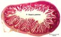
Intetsine w/Peyer's patch
©RVC 2008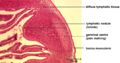
Peyer's patch
©RVC 2008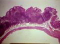
Peyer's patches
Courtesy of BioMed Archive


Function
Peyer’s patches have a similar role to that of the avian bursa of Fabricius in maturing and differentiation immature B lymphocytes. Antigens are presented to the B lymphocytes in the follicle which causes the B cells to become committed to IgA synthesis. In ruminants and pigs Peyer's patches in the ileum have a primary lympoid fuction while those in the jejenum have a secondary lymphoid function.
In pathology
- Septicaemic salmonellosis affects the Peyer's patches
- Bovine Viral Diarrhoea disease causes ulcers to form over the patches
- Infected by mycobacteria in Johnes disease
- Yersinia enter the intestinal mucosa via M cells
| Originally funded by the RVC Jim Bee Award 2007 |
Error in widget FBRecommend: unable to write file /var/www/wikivet.net/extensions/Widgets/compiled_templates/wrt662e5f2361beb1_01075286 Error in widget google+: unable to write file /var/www/wikivet.net/extensions/Widgets/compiled_templates/wrt662e5f236561f6_24564149 Error in widget TwitterTweet: unable to write file /var/www/wikivet.net/extensions/Widgets/compiled_templates/wrt662e5f23692856_67109293
|
| WikiVet® Introduction - Help WikiVet - Report a Problem |