CNS Vasculature - Anatomy & Physiology
Circle of Willis
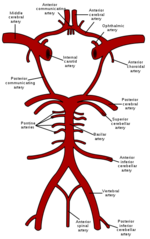
Blood is supplied to the brain from a ventral arterial supply in all species; from a circle of arteries called the Circle of Willis (also called the cerebral arterial circle or arterial circle of Willis) which lies ventrally to the hypothalamus where it forms a loose ring around the infundibular stalk. Although the appearance of the circle of Willis is fairly constant amongst mammals, the sources of blood supply to the circle and the direction of flow around the circle are very species specific. Blood is supplied to the brain by the internal carotid artery in dogs and horses whilst in other domestic species the main blood supply is from branches of the maxillary artery. The Circle of Willis is made up of five main pairs of vessels;
- Rostral Cerebral Arteries: supply the medial aspect of the cerebral hemispheres.
- Middle Cerebral Arteries: supply the lateral and ventrolateral aspects of the cerebral hemispheres.
- Caudal Cerebral Arteries: supply the occipital lobes.
- Rostral Cerebellar Arteries: supply the rostral aspects of the cerebellum
- Caudal Cerebellar Arteries: supply the caudal and lateral aspects of the cerebellum.
The arrangement of the Circle of Willis means that if one part of the circle becomes blocked or narrowed (stenosed), or one of the arteries supplying the circle is stenosed, blood flow from the other blood vessels can continue to provide a continuous supply of blood to the brain.
Blood Supply to the Circle of Willis: The Basics
In order to provide details of the species differences regarding blood supply to the brain, it is necessary to highlight the main areas of anatomy that are important within and around the circle of Willis. The following information is general and is used to provide knowledge and background to the species specifics given below.
The main blood supply to the circle is via the paired internal carotid arteries and the basilar artery. The basilar artery receives blood from the ventral spinal artery and the vertebral artery (the vertebral artery is a branch of the subclavian artery running through the vertebral foramina of C1 - C6). The internal carotid artery receives blood supply from the external carotid artery, the common carotid artery and in some species also from the vertebral artery via the occipital artery. The external carotid artery itself can also receive blood from the maxillary artery. In some species the maxillary artery is also directly able to supply the internal carotid artery, bypassing the external carotid artery via an anastomising ramus linking the internal carotid and maxillary arteries. This maxillary anastomising ramus allows blood to flow through the maxillary rete mirabile which is a network of vessels located within the cavernous sinus, facilitating the cooling of blood temperature and reducing fluctuations from pulsatile blood flows (see rete mirabile section below for further details on function). As mentioned above, the vertebral artery can also supply the internal carotid artery via the occipital artery but this too can be bypassed so that the vertebral artery can directly supply the internal carotid artery via a ramus to the internal carotid directly from the vertebral artery.
Rete Mirable
The brain is particularly susceptible to increased blood temperature and to protect the brain from any potential heat stress a number of species have developed protective mechanisms with the ability to selectively cool the brain. This protective system is often referred to as the Rete Mirable.
The Rete Mirable is a complex network of arteries and veins lying very close to each other and depends on a countercurrent blood flow between the arterioles and venules (blood flowing in opposite directions). It exchanges heat, ions, or gases between vessel walls so that the two bloodstreams within the rete maintain a gradient. The image shows the countercurrent exchange in the carotid rete mirable (carotid rete) of a sheep controlling the temperature of blood supplied to the brain.
Species Differences: Blood Supply
The blood supply to the brain has major differences depending on species. Across all species, there are variations in four major potential channels to facilitate blood supply to the Circle of Willis. As mentioned above, these are;
- Internal carotid artery
- Basiliar artery
- The anastomosing ramus from the maxillary artery to the internal carotid artery
- The connection of the vertebral artery to the internal carotid artery
Dog and Man (and many other species)
The circle of Willis in the dog is supplied from three sources; paired internal carotid arteries laterally and the basilar artery caudally. The internal carotid artery is a terminal branch of the common carotid artery. The internal carotid artery blood reaches all of the cerebral hemisphere except for its most caudal part. Vertebral blood supplies the remainder of the cerebral hemisphere and the rest of the brain. Vertebral arteries are responsible for almost all supply to the occipital lobes of the cerebral hemisphere in the dog.
Sheep and Cat
Again the main supply to the brain in both species is via three main sources; paired internal carotid arteries and the basilar artery.
Where the vertebral artery supplies the occipital artery and this then supplies the internal carotid, this connection is missing so the occipital is only able to join the external carotid and the common carotid arteries. Similarly the vertebral artery ramus is also missing so there is no direct blood supply to the internal carotid from the vertebral artery and also no associated rete mirabile. The maxillary ramus supplying the internal carotid is still patent on the left side. Maxillary blood is distributed to all of the brain except the caudal part of medulla oblongata, which is supplied by vertebral blood.
Ox
As with the previous species, the blood supply to the circle of Willis is via three main routes; paired internal carotid arteries and a basilar artery. The ox is also missing a connection from the occipital artery/external carotid/common carotid to the internal cartoid in the same manner as the ovine/feline physiology. However, where the ovine/feline physiology included only a maxillary ramus, the ox has maxillary and vertebral rami, with associated rete mirabile. Therefore the ox has four functional rete mirabile involved in blood supply to the circle of Willis. A mixture of maxillary and vertebral blood reaches all parts of the brain.
In the Ox the maxillary rami containing the rete mirabile are associated with the cavity of the frontal sinus which helps to facilitate cooling of the blood.
Species Differences: Regions of Blood Supply
Dog and Man
In dog and man the internal carotid artery supplies approximately 3/4 of the cerebrum whilst the vertebral artery supplies the very caudal aspect of the cerebrum, the cerebellum, medulla oblongata and brain stem.
Sheep and Cat
Maxillary blood (via the maxillary rami) supplies the whole of the cerebrum, the cerebellum and the medulla oblongata. Vertebral blood supplies structures caudal to the cerebellum and the brain stem.
Ox
Due to the higher number of rami in the ox, maxillary and vertebral blood are able to supply the whole brain with no clear distinctions in supply area as seen in the species above.
| This article has been peer reviewed but is awaiting expert review. If you would like to help with this, please see more information about expert reviewing. |


