Cow Mammary Gland - Anatomy & Physiology
General Structure
The mammary gland of the cow takes on added significance due to the importance of milk as a human food source. The mammary gland comprises four mammary complexes, which are separate units, consolidated in a single mass - the udder. The udder hangs from the caudal abdomen and the pelvis. The quarters are all four parts of the bovine udder, each associated with one teat. All four quarters are completely separated from each other. Accessory teats are sometimes associated with functional glandular tissue, although they are undesirable and may complicate milking if they are fused or too close to the principle teats.
Appearance
Varies greatly with breed.
Blood Supply
Arteries
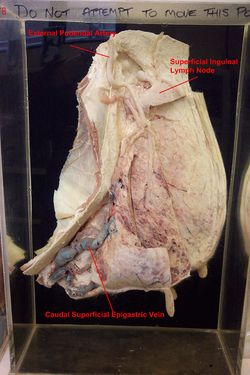
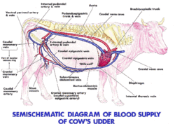
The main artery to the udder is a direct continuation of the external pudendal artery. It enters the base of the udder on its dorsocaudal aspect, after passing through the inguinal canal. It forms a sigmoid flexure before dividing into a cranial and caudal mammary artery. The sigmoid flexure permits the artery to stretch when the udder is full of milk and thus heavier than usual. Mammary arteries anastamose with the superficial caudal epigastric artery, which is connected to the cranial epigastric artery. Due to the requirement for extensive blood supply during lactation, there is a small contribution from the internal pudendal artery.
Veins
Drainage from the udder is through; 1. external pudendal vein
Drains through the inguinal canal. It also has a sigmoid flexure to allow stretching when the udder fills.
2. Superficial cranial epigastric veins
The superficial cranial and caudal epigastric veins anastamose to form the large abdominal 'milk vein' on the ventral abdomen. Incompetent valves allow blood to flow in either direction. This is important for maintaining drainage of the extensive amount of blood present. For example, if the cow were to lay down, the thin walled veins on that side would be easily occluded. The milk vein passes through the abdominal wall, caudal to the costal arch to join the internal thoracic vein, known as the 'milk well'.
3. Furstenburg's venous ring
Furstenburg's venous ring are the venous circle at the junction of the teat and gland sinuses.


