Egg Composition and Formation - Anatomy & Physiology

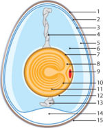
Schematic Diagram of a Chicken Egg: (1. Eggshell 2. Outer membrane 3. Inner membrane 4. Chalaza 5. Exterior albumen (outer thin albumen) 6. Middle albumen (inner thick albumen) 7. Vitelline membrane 8. Nucleus 9. Germinal disk (blastoderm) 10. Yellow yolk 11. White yolk 12. Internal albumen 13. Chalaza 14. Air cell 15. Cuticle)
©RVC 2008Size
- Varies greatly between species.
- Altricial species lay much smaller eggs than Precocial species.
- Precocial: Chicks have natal down,hatch with their eyes open and can survive outside the nest within 1-2 days.
- Altricial: Chicks are born blind and nakes,require long periods of feeding.
Shell
The shell provides physical protection. It protects the embryo from microorganisms and regulates evaporation so that the embryo does not become dessicated. The shell is also a source of calcium carbonate, which is important for bone formation in the embryo.
Shell membranes
- Organised into an inner and outer layer which lie in close apposition.
- Part at one end to form the air cell, where the hatching chick draws its first breath.
- Meshwork of protein fibres (protein and glycoprotein) with minor constituents collagen, hydroxyproline and hydroxylysine.
- Prevent dessication and infection.
Testa
- Main thickness of the shell
- Matrix fibres and calcium carbonate
- Calcium carbonate layes contains pores to allow gas exchange.
- Pigmented by porphyrin and biliverdin
Cuticle
- Water repellent
- Barrier to infection
Yolk
- Protein and lipids are synthesized in the liver and travel to the oocyte in the ovary, where they are made into yolk (vitellogenesis).
- Principle stored nutrients, provides main nutrient for the embryo.
- Produces water and energy, allowing survival in arid environments.
- Thick and viscous
- Contains maternal antibodies, primarily IgG.
Albumin
- Less viscous than yolk
- Composed mainly of water and protein.
- Buffers the embryo from sudden changes in temperature.
- Serves as a shock-absorber to protect the embryo.
- Thin layer encloses the yolk membranes and suspends the yolk in the centre of the egg by twisted strands called Chalazae.
Germinal Disc
- Small, circular, white spot on the surface of the yolk.
- Contains cytoplasm and the oocyte.
Embryonic Membranes
Within the egg, three extraembryonic membranes support the life & growth of the embryo:
Amnion
- Surrounds only the embryo
- Inner layer of cells secretes amniotic fluid in which the embryo floats.
- Fluid keeps the embryo from drying out and provides protection.
Chorion
- Surrounds all embryonic structures and serves as a protective membrane (chorioallantoic membrane).
- Also important for:
- Transpiration
- Metabolism
- Waste collection
- Calcium transfer from the shell to the embryo.
Allantois (or allantoic sac)
- Grows larger as embryo grows
- Fuses with the chorion and is called the chorio-allantoic membrane.
- Works together with chorion to permit respiration (exchange of oxygen and carbon dioxide) and excretion.
- Important in storage of nitrogenous wastes (uric acid).