File:Giardiasis duodenum high power.jpg
Giardiasis_duodenum_high_power.jpg (800 × 533 pixels, file size: 118 KB, MIME type: image/jpeg)
Summary
| Description |
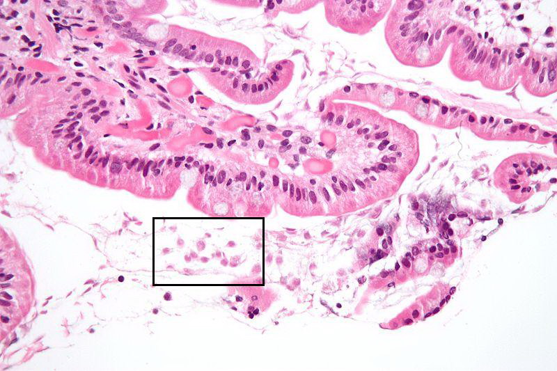
High magnification micrograph of a duodenal mucosal biopsy with giardiasis, H&E stain. The Giardia organisms are seen in the bowel lumen (bottom of image, boxed) and are: * Pale staining/translucent. * Size: 12-15 micrometers (long axis) x 6-10 micrometers (short axis). * Mostly seen to be semicircular or crescent shaped, as the long axis of the organism is rarely in the plane of the (histologic) section. The villi have interepithelial lymphocytes, but have a normal architecture. |
|---|---|
| Date |
Originally uploaded 15th July 2009 |
| Source |
Wikimedia Commons |
| Author |
Nephron |
| Permission (Reusing this file) |
Permission is granted to copy, distribute and/or modify this document under the terms of the GNU Free Documentation License, Version 1.2 or any later version published by the Free Software Foundation; with no Invariant Sections, no Front-Cover Texts, and no Back-Cover Texts. |
Licensing
| This file is licensed under the Creative Commons Attribution-Share Alike 3.0 Unported license. | |
|
File history
Click on a date/time to view the file as it appeared at that time.
| Date/Time | Thumbnail | Dimensions | User | Comment | |
|---|---|---|---|---|---|
| current | 18:35, 11 August 2010 | | 800 × 533 (118 KB) | Lizzies (talk | contribs) | {{Information |Description=High magnification micrograph of a duodenal mucosal biopsy with giardiasis, H&E stain. The Giardia organisms are seen in the bowel lumen (bottom of image, boxed) and are: * Pale staining/translucent. * Size: 12-15 microm |
You cannot overwrite this file.
File usage
The following page uses this file: