Ornamental Fish Q&A 16
| This question was provided by Manson Publishing as part of the OVAL Project. See more Ornamental Fish Q&A. |
You are preparing a koi for surgery to remove an abdominal mass. Your anesthetic protocol is established and you plan on a mid ventral incision with the fish in dorsal recumbency.
| Question | Answer | Article | |
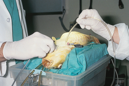
| How will you monitor the fish while it is anesthetized? | Opercular movement is a good indicator of life, but in cases where the fish needs to be very deep, opercular movements may cease. Electrocardiography is an option, with leads placed at the base of each pectoral fin and one near the vent. The alligator clips can be attached to small needles placed in the skin. |
Link to Article | |
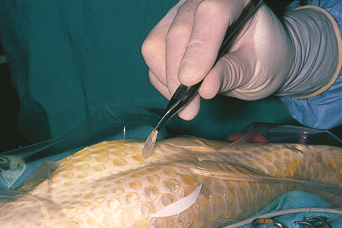
| How will you prepare the incision site? | A clear sterile surgical drape should be used and the scales removed along the planned incision line. The skin can be treated carefully with a pre-surgical antiseptic, although this may not be necessary. |
Link to Article | |
| How will you manage the case post-operatively? | The mass turned out to be an ovarian granulosa-theca cell tumor and it was removed uneventfully. The skin was closed with nonabsorbable nylon suture and air was aspirated from the abdominal compartment following surgical closure. Note the use of an extra plastic tube to provide fresh water to the gills (two tubes providing water flow are in the mouth of the fish). Enrofloxacin was administered intramuscularly at a dose of 10 mg/kg every 48 hours for ten days. This fish recovered and was doing well five months after surgery. |
Link to Article | |

