Small Animal Abdominal and Metabolic Disorders Q&A 05
| This question was provided by Manson Publishing as part of the OVAL Project. See more Small Animal Abdominal and Metabolic Disorders Q&A. |
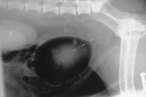
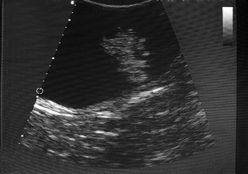
A seven and a half-yearold, entire male Cocker Spaniel was presented with haematuria of several months’ duration. The blood was always mixed with urine and tended to be worst towards the end of urination. Radiographic and ultrasonographic investigations revealed the lesions shown.
| Question | Answer | Article | |
| What is your main differential diagnosis? | The main differential diagnosis is polypoid cystitis; this is based upon the size, position and number of the intravesicular masses. On ultrasound examination of the bladder the polyps do not invade the bladder wall but appear to arise from the urothelium. However, the diagnosis should be confirmed histopathologically. |
Link to Article | |
| How would you treat this animal, and what complications may occur? | Any underlying cause for the polypoid cystitis should be sought and treated, e.g. calculi, urinary tract infection. Polypoid cystitis is a poorly defined condition but is thought to occur secondary to chronic bladder irritation. Animals may show signs of haematuria and occasionally urinary frequency, but may also be asymptomatic. Treatment may be medical or surgical. In some cases the polyps may resolve spontaneously following treatment of a urinary tract infection or dissolution of urinary calculi. However, in persistent cases or where cystic calculi require removal the diagnosis is usually confirmed and the animal treated concomitantly by performing excisional biopsies of the masses. A ventral cystotomy allows good access to the bladder lumen. The bladder is everted and each polyp isolated by applying some mosquito forceps at the base of the polyp. The polyp is excised and the forceps left in place for several minutes until haemostasis is achieved. This technique absolves the use of suture material within the bladder lumen. The main complication of the surgery is haemorrhage. This is usually minor and self-limiting; however, if severe haemorrhage occurs during recovery, a clot may form within the bladder lumen. This may obstruct the bladder neck, like a ball valve, causing postoperative obstruction or dysuria. The risk of this occurring may be reduced by encouraging frequent urination and creating a diuresis by administering intravenous fluids. |
Link to Article | |

