Small Animal Soft Tissue Surgery Q&A 06
| This question was provided by Manson Publishing as part of the OVAL Project. See more Small Animal Soft Tissue Surgery Q&A. |
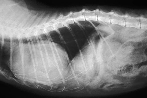
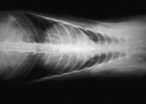
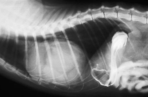
You are presented with a two-year-old, spayed female Cocker Spaniel with a history of lethargy and anorexia. Survey thoracic radiographs are shown.
| Question | Answer | Article | |
| Describe the radiographic findings. | The cardiac silhouette is enlarged and contains opacities consistent with soft tissue and fat. The trachea is displaced dorsally. The liver shadow is not identified in the abdomen and loops of gas-filled intestine are in contact with the diaphragm. The diaphragm becomes indistinct ventrally. |
Link to Article | |
| What is the most likely diagnosis? | Peritoneopericardial diaphragmatic hernia. |
Link to Article | |
| How could you confirm this diagnosis? | Echocardiography is best to confirm the diagnosis by imaging abdominal viscera such as spleen and liver within the pericardial sac. An upper gastrointestinal contrast radiographic study could also be used. In this case contrast approached the diaphragm but did not enter the pericardial sac; however, intestinal proximity to the diaphragm confirms the diagnosis, as normally the liver is interposed between these structures. |
Link to Article | |
| Describe the pathophysiology of this condition. | Peritoneopericardial diaphragmatic hernia is usually congenital resulting from incomplete closure of the diaphragmatic septum transversum. Although present at birth, it often does not cause clinical signs until later in life and may be discovered incidentally. This defect is considered a developmental, not genetic or inherited, defect. Symptomatic patients generally have signs referable to the cardiopulmonary or gastrointestinal systems. |
Link to Article | |
| Describe the surgical treatment of this condition. | The hernia is accessed by ventral midline celiotomy. To reduce the hernia, the defect may need to be enlarged as the contents may be swollen. Adhesion of the hernia contents to the pericardium or epicardium is rare. Once reduced, the defect is closed using interrupted or continuous sutures with an absorbable material. Dorsal (deepest) sutures are placed first with closure continuing ventrally. Single layer closure is sufficient and it is not necessary to separate the pericardium from the peritoneum. Air is removed from the pericardial sac by aspiration though the suture line. This is especially important when the hernial sac is large and pneumopericardium could impair pulmonary expansion. |
Link to Article | |
| What is the long-term prognosis following surgery for this patient? | Prognosis following herniorrhaphy is excellent. |
Link to Article | |


