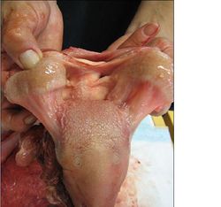
Tongue - Anatomy & Physiology
Introduction
The tongue (lingua) occupies the ventral aspect of the oral cavity and oropharynx. It is involved with grooming, lapping, prehension and manipulating food in the oral cavity. It is also involved in the deglutition reflex and vocalisation. The tongue is capable of vigorous and precise movements due to the apex being free of attachments to the oral cavity.
Structure and Function
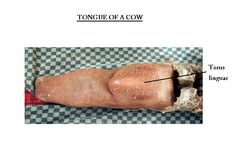
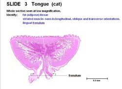
The tongue is skeletal muscle dorsally and structural fat surrounded by a cartilagenous sheath forming lyssa (canids only) ventrally. It has an attached root and body with a free apex. The frenulum (fold of mucosa) attaches the body of the tongue to the floor of the oral cavity. The root of tongue is attached to the hyoid bone. In the horse and dog, the tongue is 'u' shaped, becoming broader towards the tip. The furrow in the centre of the canid tongue is called the median sulcus. In the ox, sheep and pig the tongue is 'v' shaped with a pointed apex. The torus linguae is a swelling across the tongue laterally which pushes food against the hard palate.
Muscles
Intrinsic Muscles
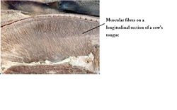
Intrinsic muscles include the dorsal and ventral longitudinal muscles and the transverse and vertical bundles.
Extrinsic Muscles
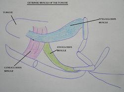
The extrinsic muscles include:
Styloglossus
Its origin is at the hyoid apparatus (stylohyoid) and it retracts and elevates the tongue.
Genioglossus
The origin is at the incisive part of the mandible. It protrudes and depressed the tongue.
Hyoglossus
The origin is at the hyoid apparatus (basihyoid). It retracts and depresses the tongue.
Geniohyoideus
It originates at the incisive part of the mandible and the insertion site is the body of the hyoid. It lies below the tongue (not within it) and draws the hyoid and therefore the tongue forward.
Innervation
All muscles moving the tongue are innervated by the hypoglossal nerve (CN XII). The rostral 2/3 of the tongue is innervated by the sensory lingual branch of the trigeminal (CN V) transmitting temperature, touch and pain sensation. The chorda tympani of the facial nerve (CN VII) transmits the taste. The caudal 1/3 of the tongue is innervated by the glossopharyngeal (CN IX) providing sensory function for taste.
Vasculature
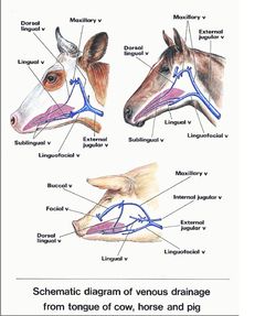
The main blood supply to the tongue is via the lingual artery, a branch of the external carotid artery. A secondary blood supply to the tongue is provided via the tonsillar branch of the facial artery and the ascending pharyngeal artery.
Histology
The tongue consists of stratified squamous epithelium. There are lingual glands and a mucosal covering tightly adheres to the contact surface. The degree of keratinisation depends on the diet. There is less keratinisation on the ventral surface and sides of tongue. It is covered by papillae for protection and taste. Papillae are specialised projections of the mucosa. Some papillae have taste buds, others are mechanical to roughen the surface of the tongue.
Types of Papillae
Conical
Conical papillae are not found in horses. They are present in the caudal 1/3 of the tongue. They point caudally and have no taste buds. There is a thick epithelium.
Foliate
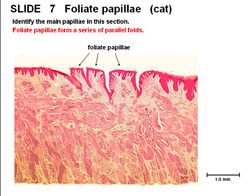
Eight to twelve papillae in parallel folds, one either side of the tongue midline. Consists of a stratified squamous epithelium, present in the caudal third of the tongue. There are taste buds, glands and lymphatics present.
Vallate
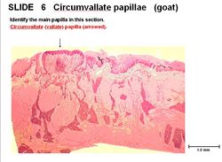
There are three to six, often secondary papillae in taste buds. There are broad glands in the caudal 1/3 of tongue. Taste buds and lymphatics are present.
Fungiform
They form the red dots on tongue surface and consist of keratinised, stratified squamous epithelium and blood vessels. They are involved in loss of heat via panting in dogs. They are present in the rostral 2/3 of the tongue and contain taste buds.
Filiform
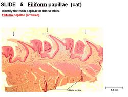
Filiform papillae are the most numerous and point caudally. There are no taste buds, glands or lymphatics. They are the smallest and consist of a thick keratin on stratified squamous epithelium. They are very prominent in cat and are present in the rostral 2/3 of the tongue.
Taste Buds
Also found on the soft palate and the pharynx (but sparsely distributed). There is a constant cell turnover, with flat, thick cells. There are taste hairs (microvilli) pointing through the taste pore. Nerves transduce chemical signals into nervous signals.
Species Differences
Canine
There are stretch receptors in the tongue and they use the tongue to lose heat by panting.
Ruminant
The tongue is heavily keratinised with long papillae for eating (protective surface). The ox has lenticular papillae which are hard and horny due to heavy keratinisation.
Feline
Feline species have long papillae for grooming, so their tongue is rough.
Porcine
Most of the papillae are soft, long and directed caudally.
Avian
The avian tongue contains a bone and is mainly used for manipulation of food rather than vocalisation like in mammals. Parrots use the tongue to produce human sounds (see species differences in syrinx)
Links
Click here for pathology of the tongue information.
| Tongue - Anatomy & Physiology Learning Resources | |
|---|---|
To reach the Vetstream content, please select |
Canis, Felis, Lapis or Equis |
 Test your knowledge using flashcard type questions |
Tongue Anatomy & Physiology Flashcards Facial Muscles |
 Selection of relevant videos |
Ventral muscles of the head potcast Lateral surface of the head of the dog potcast Lateral surface and sagittal section of the head of a sheep Lateral surface of the head of the dog potcast 5 |
 Selection of relevant PowerPoint tutorials |
Histology of the oral cavity, see part 1 for the tongue |
Anatomy Museum Resources |
Image - Cat Tongue |
Webinars
Failed to load RSS feed from https://www.thewebinarvet.com/gastroenterology-and-nutrition/webinars/feed: Error parsing XML for RSS
