Difference between revisions of "Lingual Gland - Anatomy & Physiology"
Jump to navigation
Jump to search
| (2 intermediate revisions by one other user not shown) | |||
| Line 1: | Line 1: | ||
| − | |||
==Overview== | ==Overview== | ||
| − | [[Image:Serous Lingual Gland.jpg|thumb| | + | |
| − | + | [[Image:Serous Lingual Gland.jpg|thumb|right|250px|Serous Lingual Gland Histology (Mouse), from [[Oral Cavity Histology resource|oral cavity tutorial part 1, slide 31]]]] | |
| + | |||
Acini with '''cuboidal''' epithelium, '''round''' basal nuclei and cytoplasm that stains '''pink''' is a '''[[Serous Salivary Gland - Anatomy & Physiology|serous]]''' secreting lingual gland in the '''body''' of the [[Tongue - Anatomy & Physiology|tongue]]. | Acini with '''cuboidal''' epithelium, '''round''' basal nuclei and cytoplasm that stains '''pink''' is a '''[[Serous Salivary Gland - Anatomy & Physiology|serous]]''' secreting lingual gland in the '''body''' of the [[Tongue - Anatomy & Physiology|tongue]]. | ||
| − | + | [[Image:Mucous Lingual Gland.jpg|thumb|right|250px|Mucous Lingual Gland Histology (Mouse), from [[Oral Cavity Histology resource|oral cavity tutorial part 1, slide 33]]]] | |
Acini with '''flattened''' basal nuclei, cytoplasm that stains '''blue''' and has a '''foamy''' appearence is a '''[[Mucous Salivary Gland - Anatomy & Physiology|mucous]]''' secreting lingual gland in the '''root''' of the [[Tongue - Anatomy & Physiology|tongue]]. | Acini with '''flattened''' basal nuclei, cytoplasm that stains '''blue''' and has a '''foamy''' appearence is a '''[[Mucous Salivary Gland - Anatomy & Physiology|mucous]]''' secreting lingual gland in the '''root''' of the [[Tongue - Anatomy & Physiology|tongue]]. | ||
| Line 12: | Line 12: | ||
{{Template:Learning | {{Template:Learning | ||
|powerpoints = [[Oral Cavity Histology resource|Oral Cavity part 1 tutorial covers lingual glands]] | |powerpoints = [[Oral Cavity Histology resource|Oral Cavity part 1 tutorial covers lingual glands]] | ||
| − | |||
}} | }} | ||
| − | |||
[[Category:Salivary Glands - Anatomy & Physiology]] | [[Category:Salivary Glands - Anatomy & Physiology]] | ||
[[Category:To Do - AimeeHicks]] | [[Category:To Do - AimeeHicks]] | ||
Revision as of 11:14, 24 May 2011
Overview

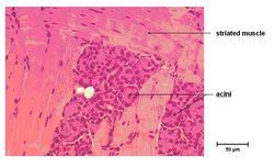
Serous Lingual Gland Histology (Mouse), from oral cavity tutorial part 1, slide 31
Acini with cuboidal epithelium, round basal nuclei and cytoplasm that stains pink is a serous secreting lingual gland in the body of the tongue.

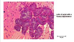
Mucous Lingual Gland Histology (Mouse), from oral cavity tutorial part 1, slide 33
Acini with flattened basal nuclei, cytoplasm that stains blue and has a foamy appearence is a mucous secreting lingual gland in the root of the tongue.
| Lingual Gland - Anatomy & Physiology Learning Resources | |
|---|---|
 Selection of relevant PowerPoint tutorials |
Oral Cavity part 1 tutorial covers lingual glands |