|
|
| Line 19: |
Line 19: |
| | }} | | }} |
| | [[Category:Histology PowerPoints]] | | [[Category:Histology PowerPoints]] |
| | + | ==User reviews:== |
| | + | ''Click 'edit' button at right side of screen to enter a user review'' |
| | + | <!---Write below this line, click 'save page' when you've finished---> |
Revision as of 11:27, 24 May 2011
Oral Cavity Histology PowerPoint tutorial (1 of 2)
Click here to access the resource.
Resource Information
| Description
|
Oral Cavity Histology resource
PowerPoint
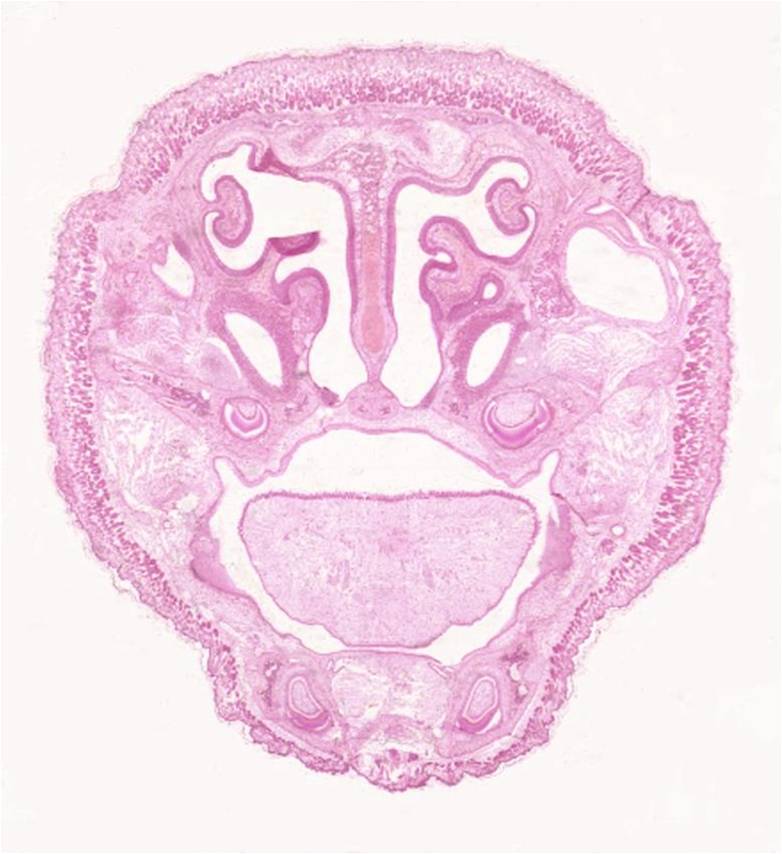
This is a tutorial on histology of the oral cavity. The PowerPoint contains many histological images of sections of the head, as well as images of different epithelial types, glands and papillae and anatomy of the region, with the opportunity for self-assessment. This also features an easily accessible menu slide, allowing rapid navigation.
This is the first of 2 oral cavity tutorials
Duration = 41 slides
|
| Date
|
2011
|
| Source
|
Royal Veterinary College
|
| Author
|
John Bredl
|
| Licensing
|
|
Oral Cavity Histology PowerPoint tutorial (2 of 2)
Click here to access the resource.
Resource Information
| Description
|
Oral Cavity Histology resource
PowerPoint
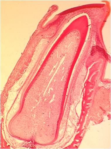
This is a tutorial on histology of the oral cavity. The PowerPoint contains many histological images of tooth development and different types of salivary glands. This also features an easily accessible menu slide, allowing rapid navigation.
This is the second of 2 oral cavity tutorials
Duration = 46 slides
|
| Date
|
2011
|
| Source
|
Royal Veterinary College
|
| Author
|
John Bredl
|
| Licensing
|
|
User reviews:
Click 'edit' button at right side of screen to enter a user review

