Difference between revisions of "Small Animal Dermatology Q&A 16"
Ggaitskell (talk | contribs) (Created page with "{{Template:Manson Moriello}} centre|500px<br> <br /> '''A 2-year-old blue doberman pinscher dog was referred for the problem of an...") |
|||
| (2 intermediate revisions by 2 users not shown) | |||
| Line 19: | Line 19: | ||
*cystic or dilated hair follicles. <br> | *cystic or dilated hair follicles. <br> | ||
As hairs regrow, they grow more slowly and are often deformed. This disorder has been seen in cats with blue or cream colored coats. | As hairs regrow, they grow more slowly and are often deformed. This disorder has been seen in cats with blue or cream colored coats. | ||
| − | |l1= | + | |l1= |
|q2=How is this disorder treated? | |q2=How is this disorder treated? | ||
|a2= | |a2= | ||
| Line 25: | Line 25: | ||
As a result, excessive grooming and harsh shampoos should be avoided. It is important to control the recurrent bacterial pyodermas associated with this disorder. <br><br> | As a result, excessive grooming and harsh shampoos should be avoided. It is important to control the recurrent bacterial pyodermas associated with this disorder. <br><br> | ||
Affected dogs will continue to lose hair, and many are alopecic by 2–3 years of age. | Affected dogs will continue to lose hair, and many are alopecic by 2–3 years of age. | ||
| − | |l2= | + | |l2= |
|q3=What are the most common histological findings? | |q3=What are the most common histological findings? | ||
|a3= | |a3= | ||
Common histological findings show melanin clumping in the epidermal and follicular basal cells, macromelanosomes in the hair shafts or hair bulbs, and follicular dysplasia. <br><br> | Common histological findings show melanin clumping in the epidermal and follicular basal cells, macromelanosomes in the hair shafts or hair bulbs, and follicular dysplasia. <br><br> | ||
The melanin clumping and macromelanosomes are key findings for diagnosis of this disease. | The melanin clumping and macromelanosomes are key findings for diagnosis of this disease. | ||
| − | |l3= | + | |l3= |
</FlashCard> | </FlashCard> | ||
Revision as of 11:34, 6 June 2011
| This question was provided by Manson Publishing as part of the OVAL Project. See more small animal dermatological questions |
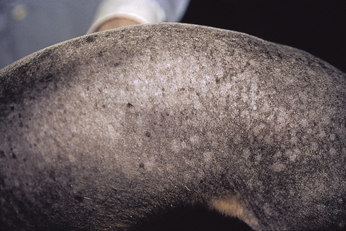
A 2-year-old blue doberman pinscher dog was referred for the problem of an ‘endocrine alopecia’ of unknown etiology. Previous diagnostic tests were normal or negative and included: skin scrapings, dermatophyte culture, complete blood count, urinalysis, serum chemistry panel, thyroid hormone evaluation, low-dose dexamethasone suppression test, and surgical neutering. Dermatological examination reveals a thin hair coat, nodular-like hair follicles, comedones, bacterial pyoderma, and scaling. All of the other littermates are similarly affected.
| Question | Answer | Article | |
| What is the most likely diagnosis, and what is the cause? | Color dilution alopecia (previously called color mutant alopecia). This is a genetic disorder of the hair coat commonly associated with a blue or fawn coat color. The cause is unknown, but it is believed to be due, in part, to a defect in the coat color genes at the D locus.
As hairs regrow, they grow more slowly and are often deformed. This disorder has been seen in cats with blue or cream colored coats. |
[[|Link to Article]] | |
| How is this disorder treated? | There is no effective treatment for this disorder. In the early stages of the disorder, hair loss is caused by hair shaft fracture. |
[[|Link to Article]] | |
| What are the most common histological findings? | Common histological findings show melanin clumping in the epidermal and follicular basal cells, macromelanosomes in the hair shafts or hair bulbs, and follicular dysplasia. |
[[|Link to Article]] | |
